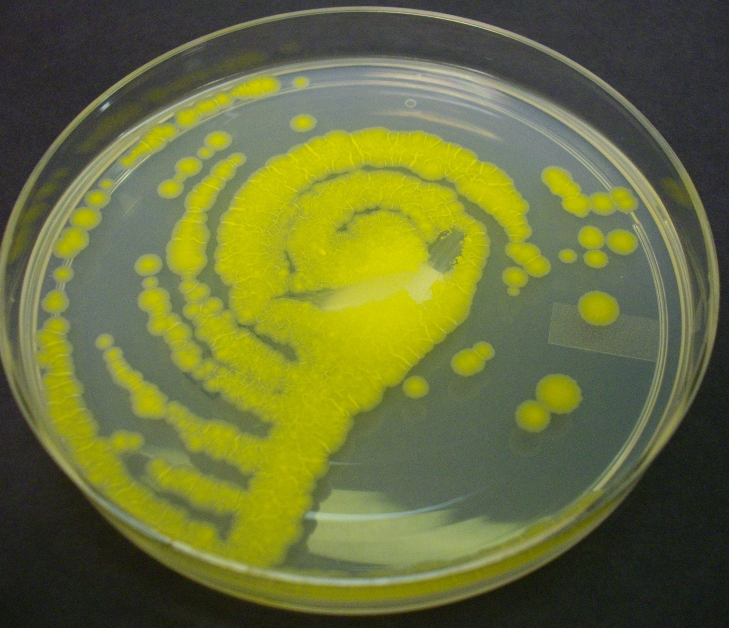
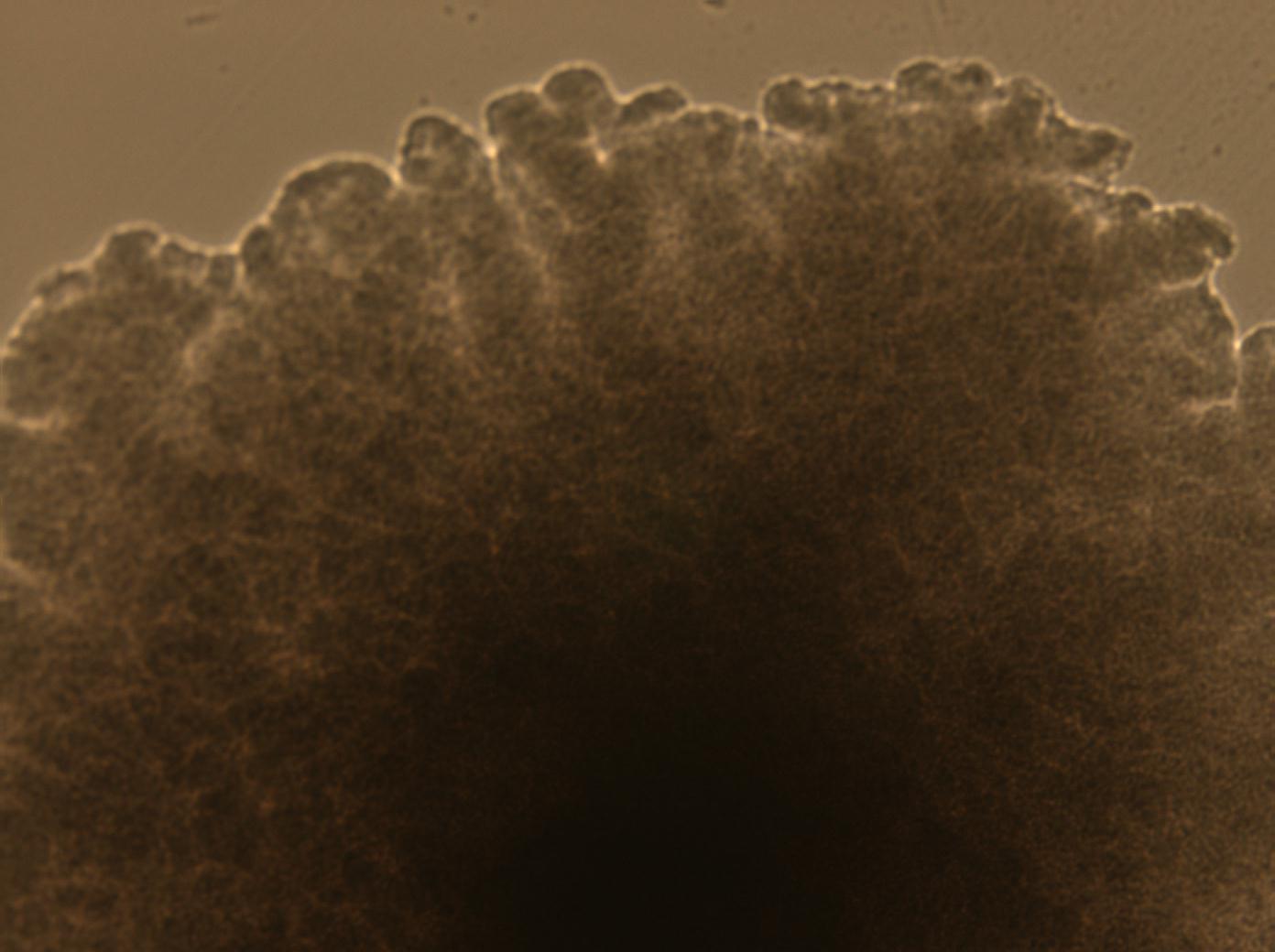
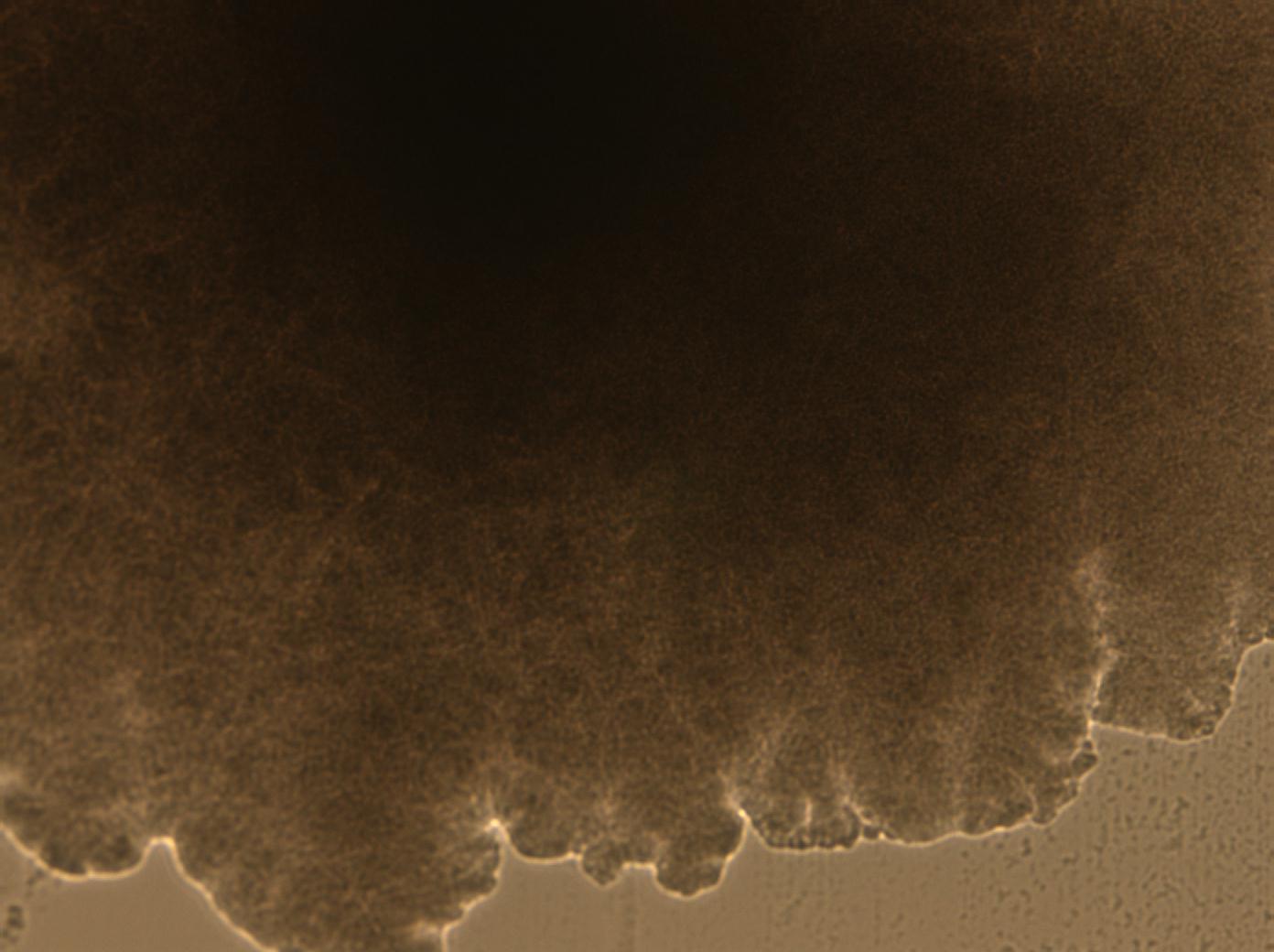
Mycobacterium kansasii is a slow-growing nontuberulous mycobacterium (NTM) found naturally in tap water systems, rivers and soil. Unlike Mycobacterium tuberculosis, it is not spread from person to person. Infection is thought to occur mainly by inhaling contaminated water aerosols or less commonly through skin exposure. It can cause disease, particularly in people with weakened immune systems or underlying lung disease.

Disease caused by Mycobacterium kansasii: Pulmonary disease is the most frequent manifestation. Chest imaging may show cavitary lesions, especially in the upper lobes, nodules, bronchiectsis and fibrosis. Lung disease often resembles tuberculosis on chest X-ray or CT scan. It often resembles pulmonary tuberculosis

Diagnosis requires combining clinical finding (symptons), Radiologic evidence (chestX-ray or CT) and Microbiology culture confirmation

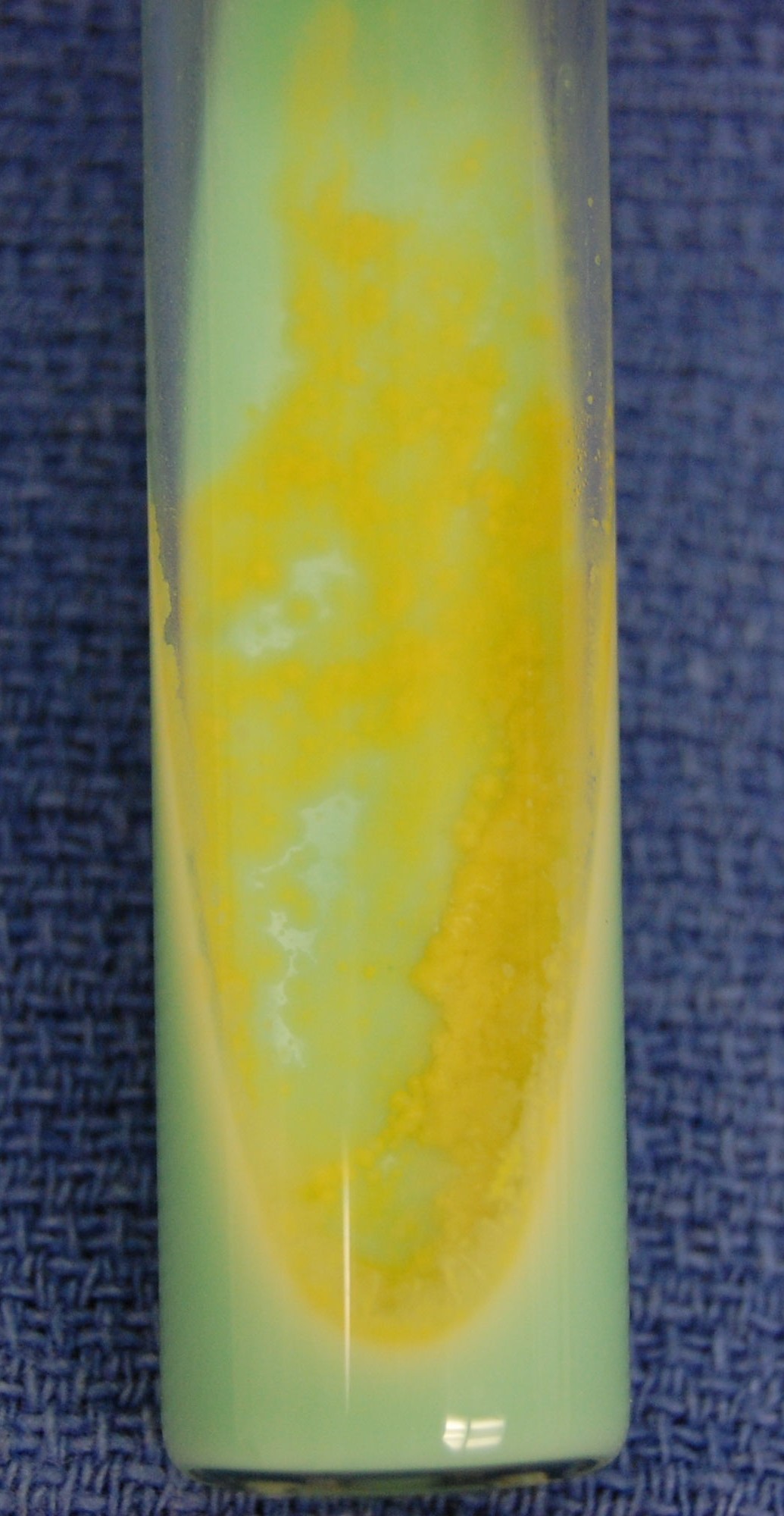
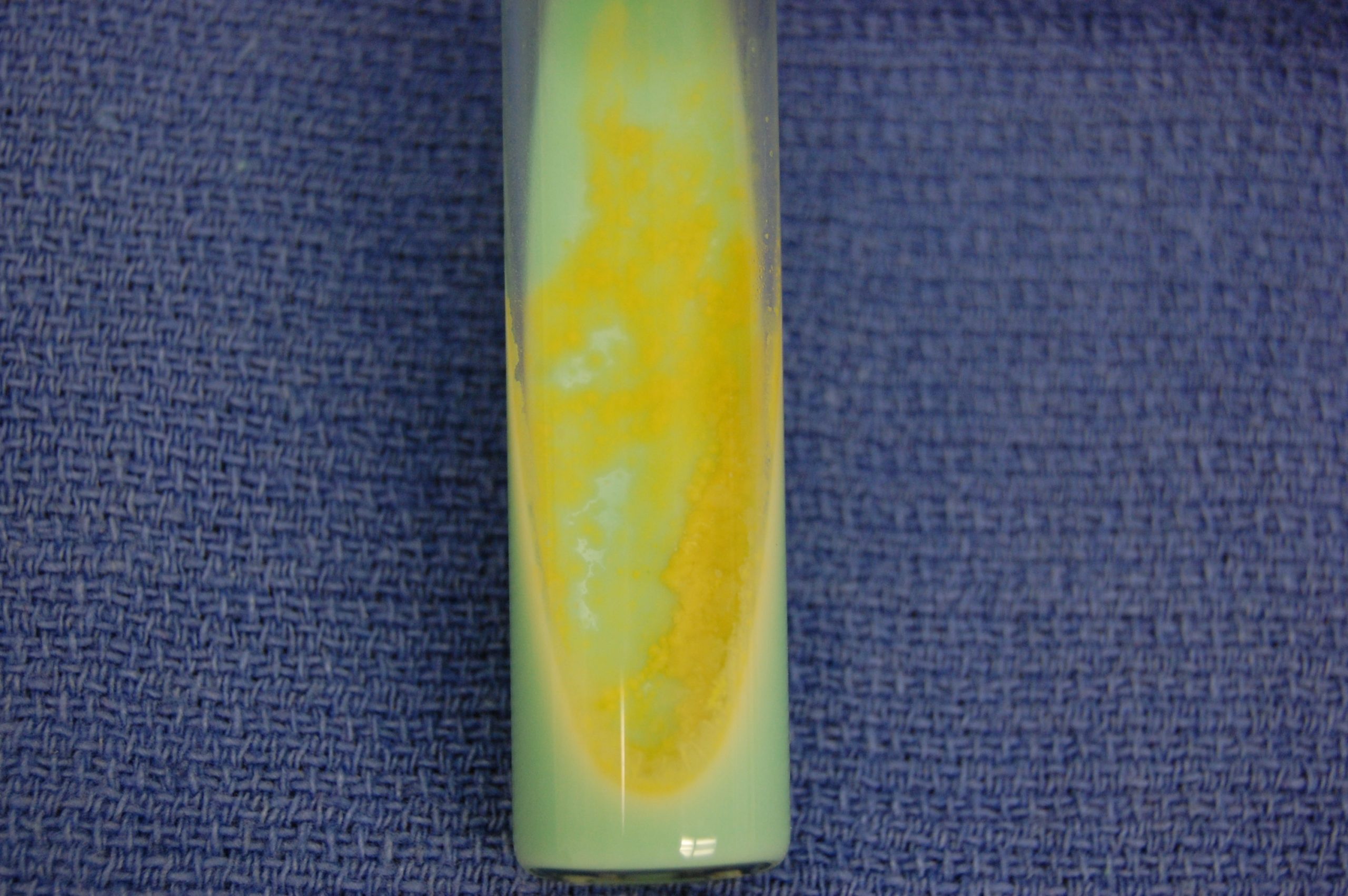
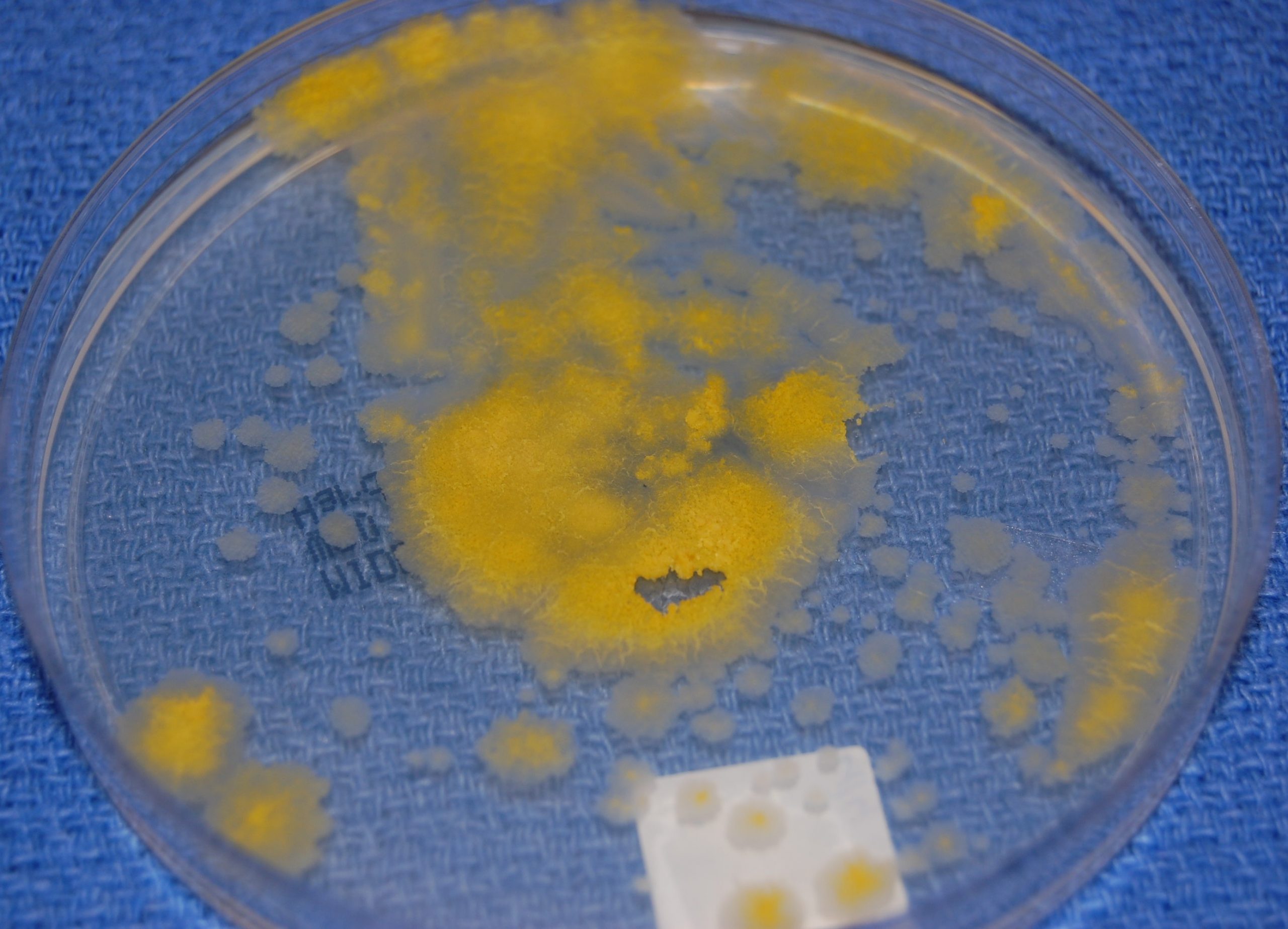
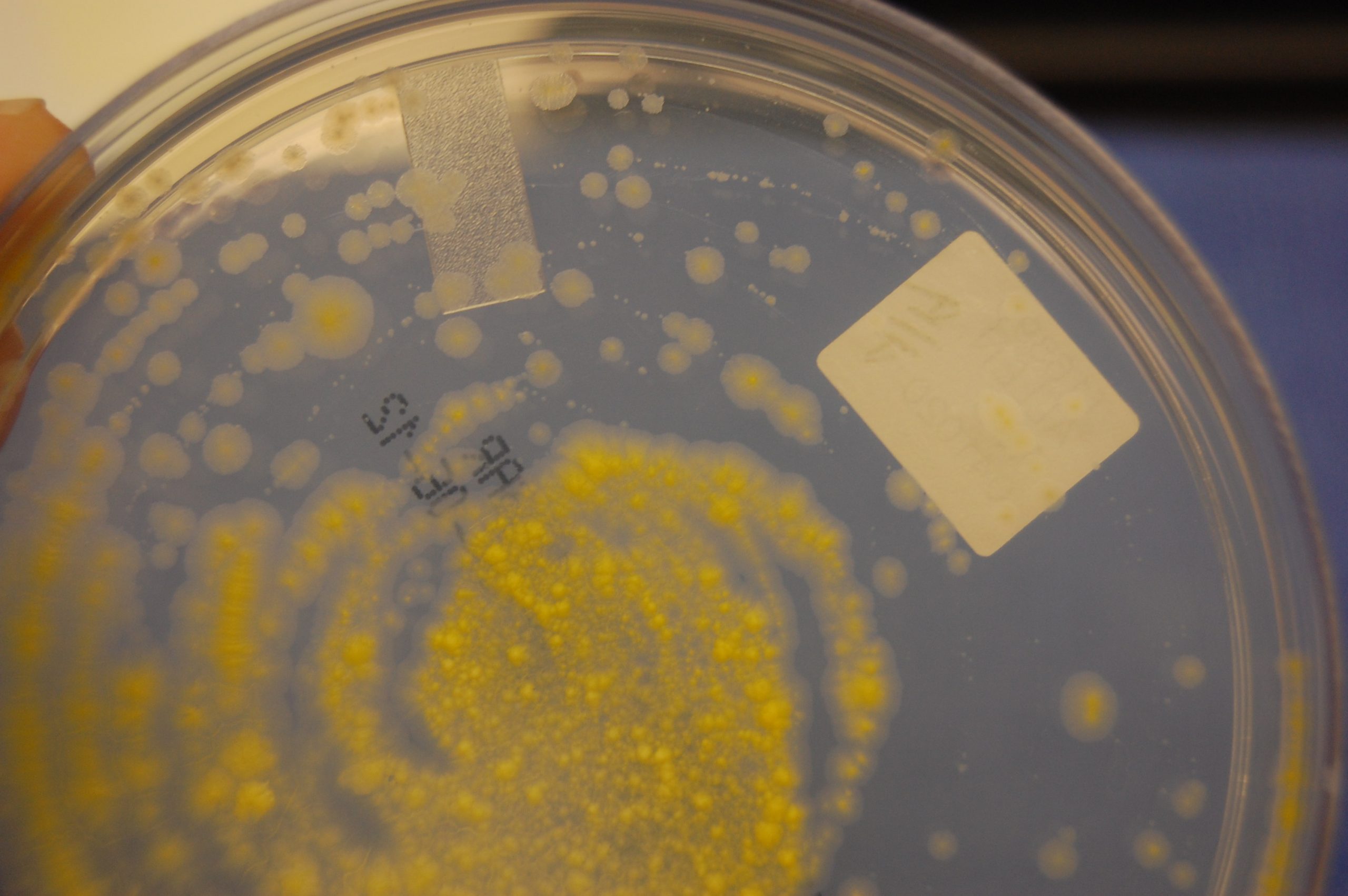
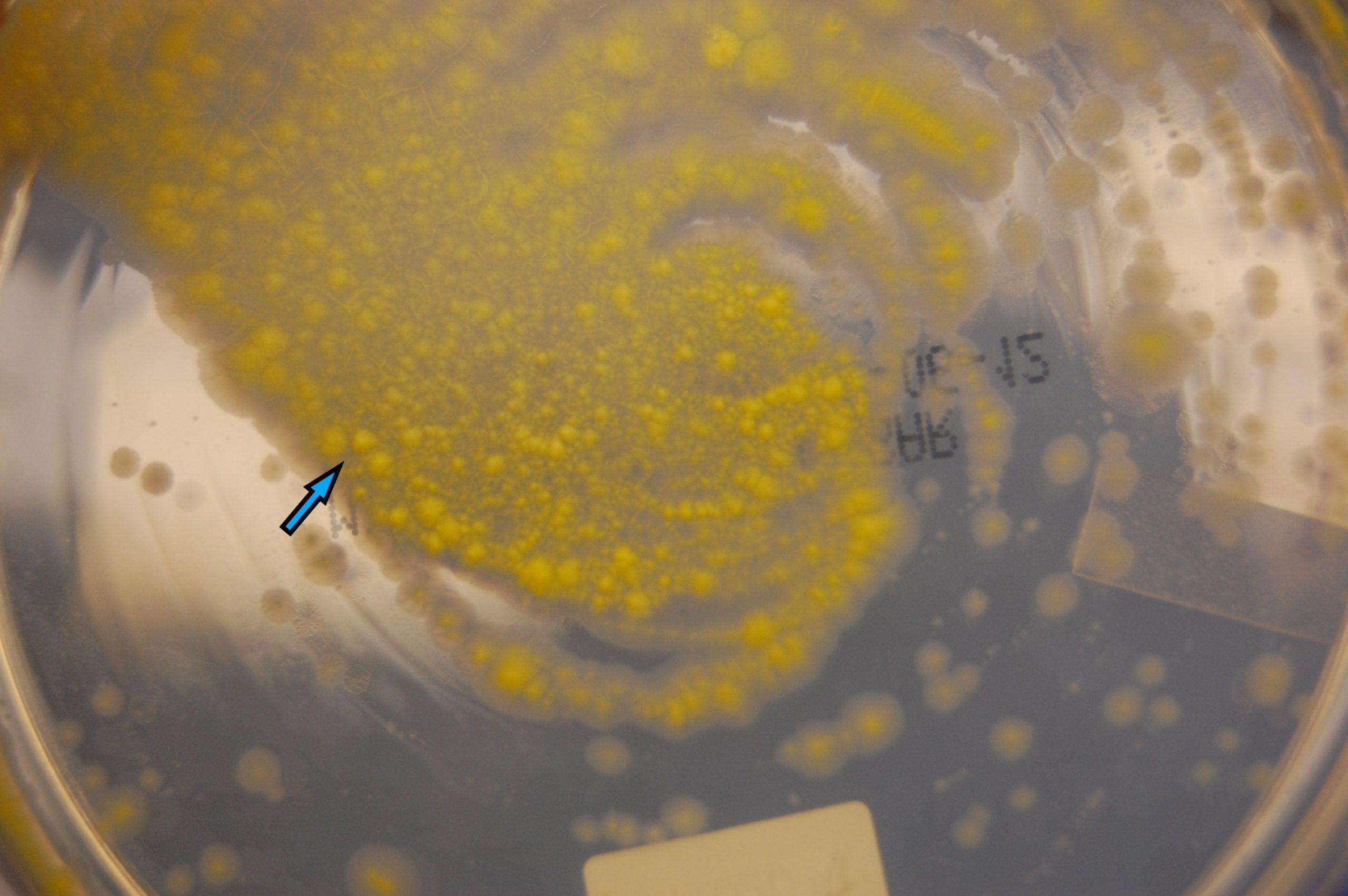
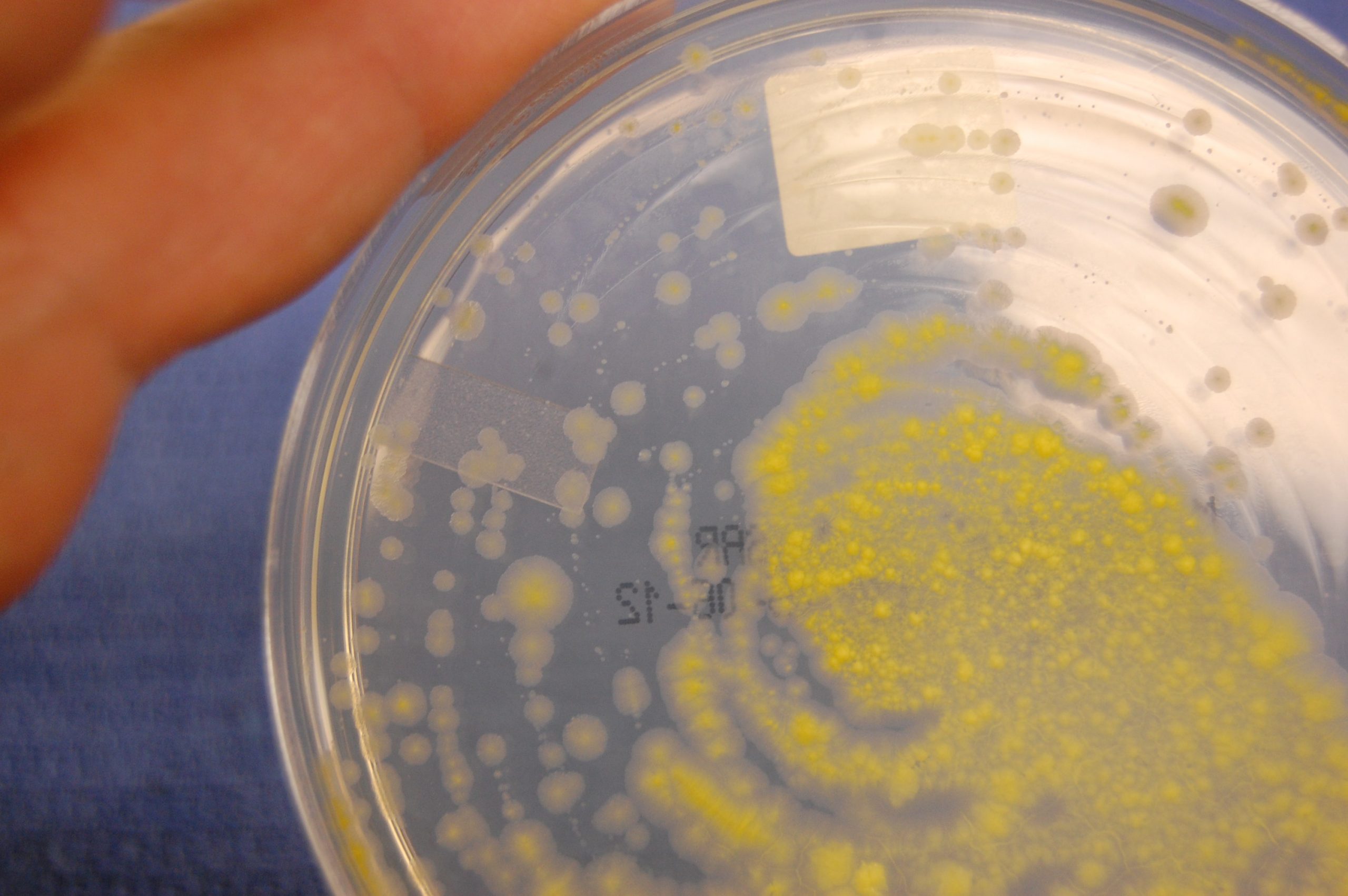
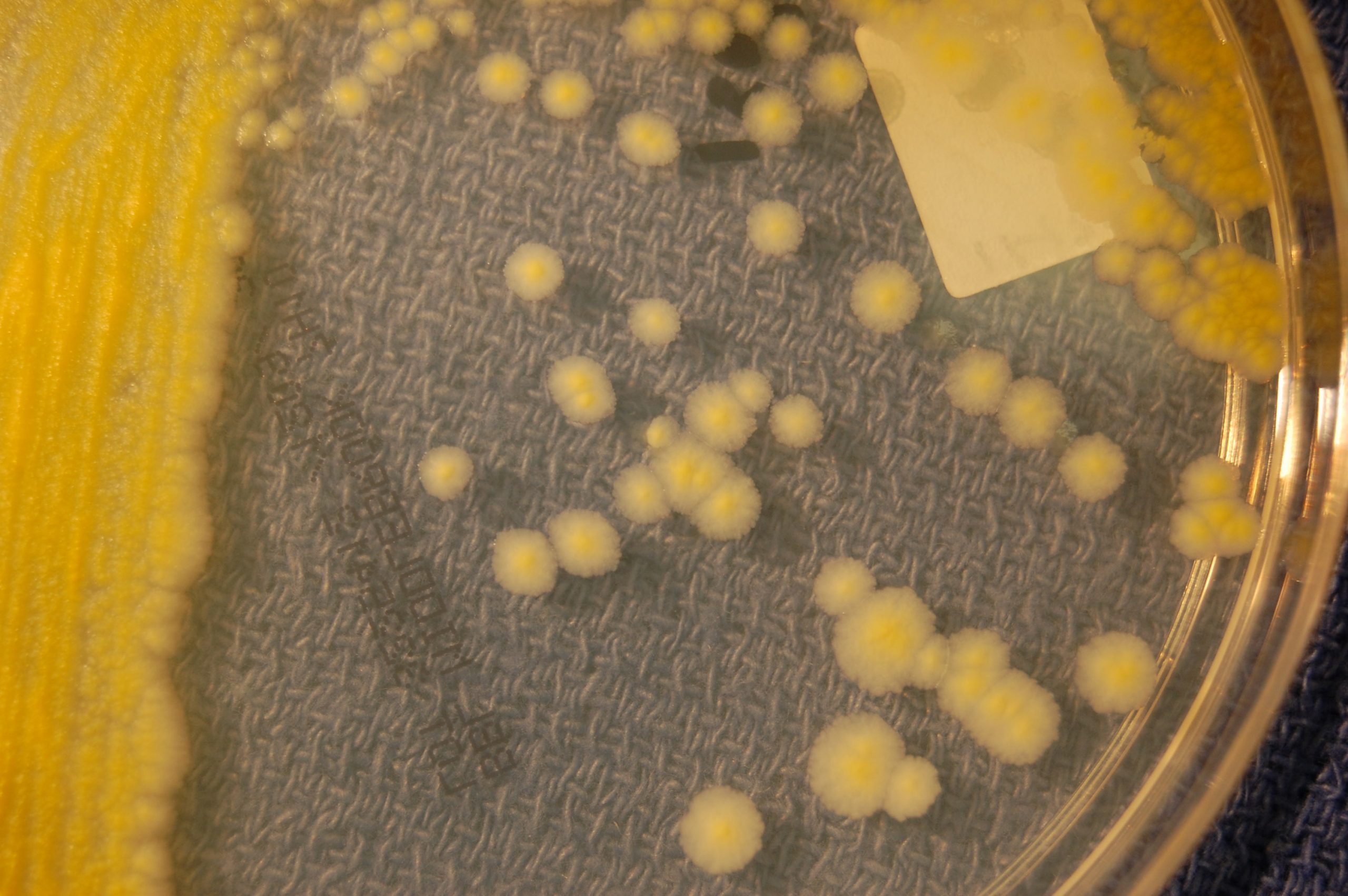
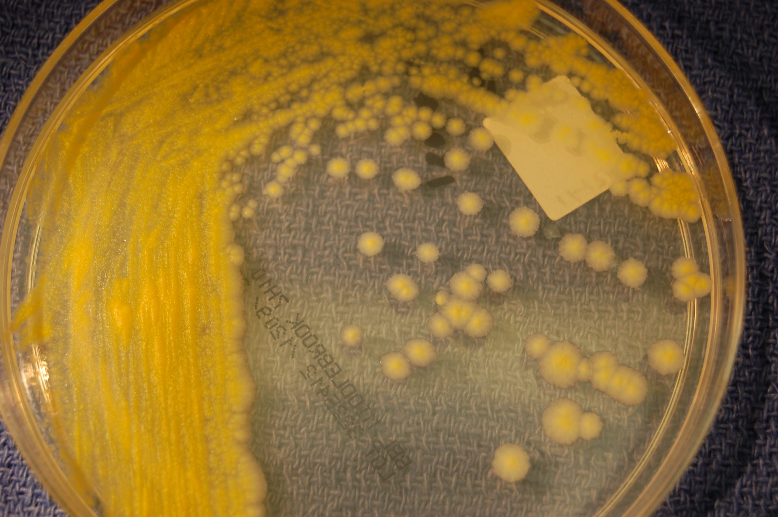
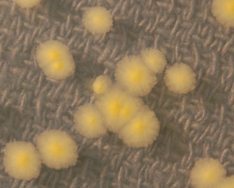
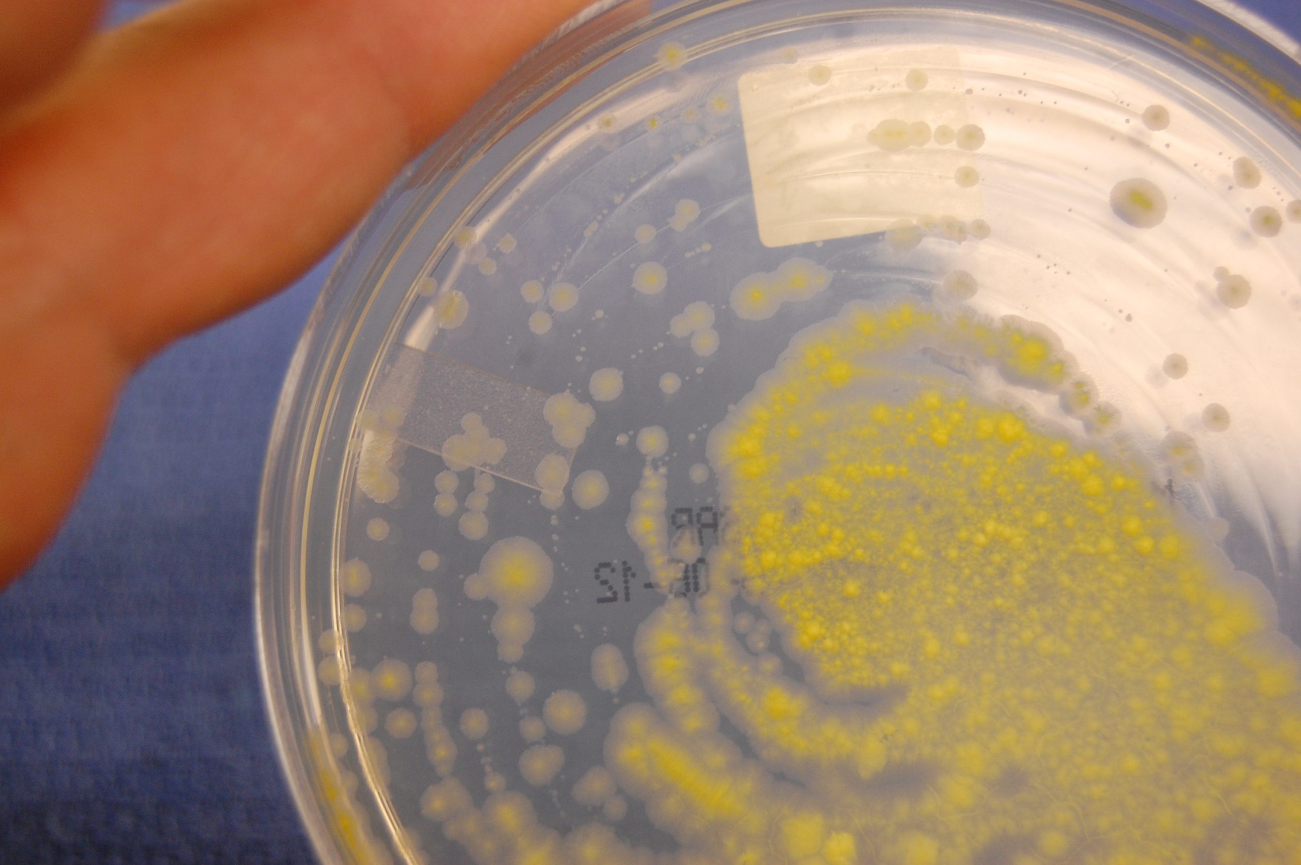
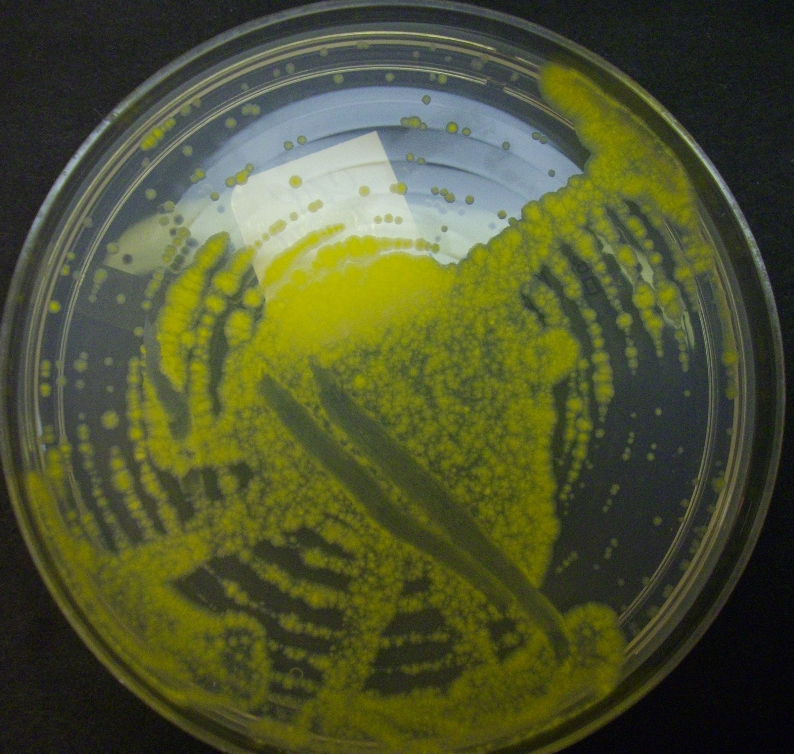
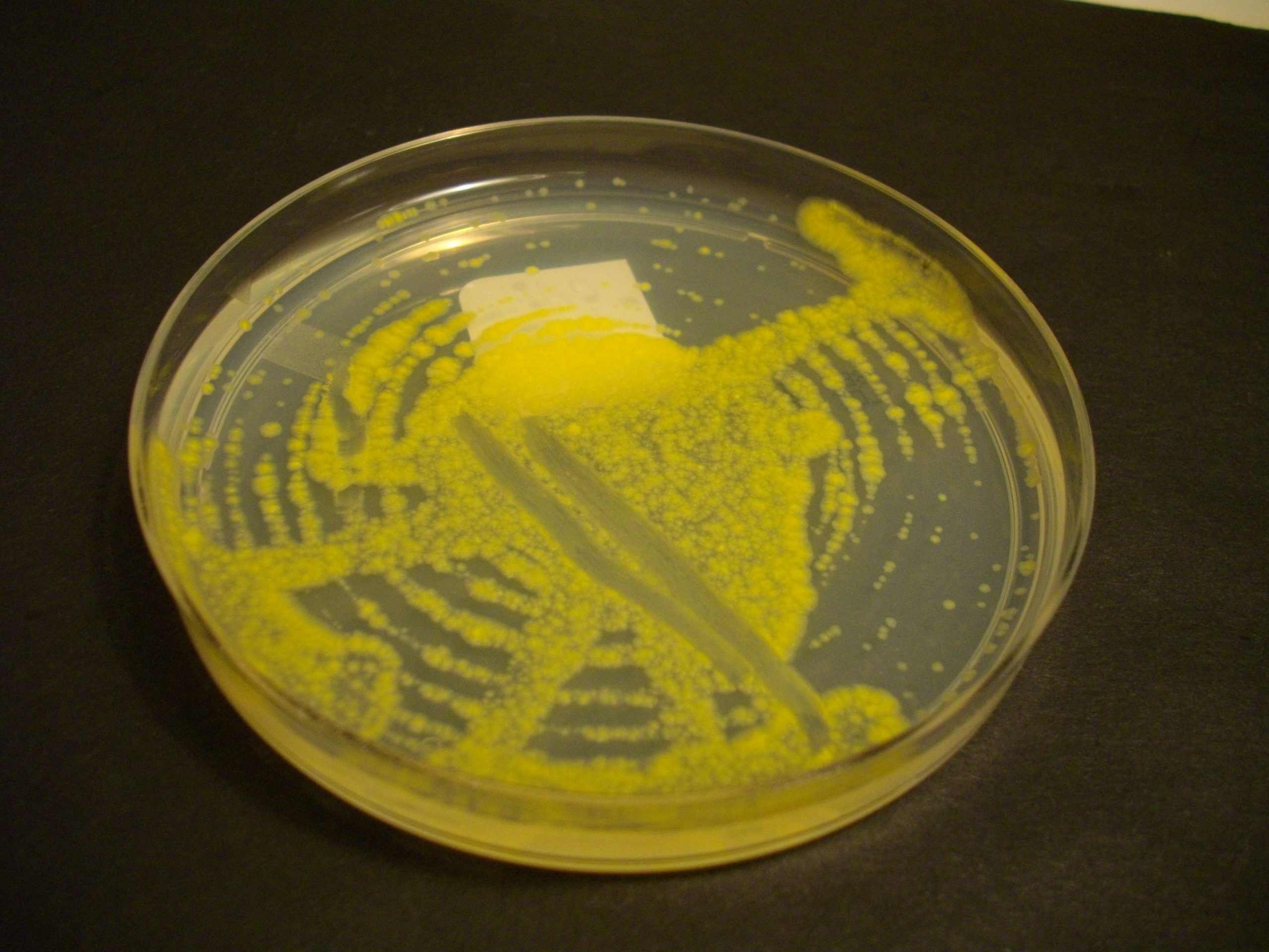
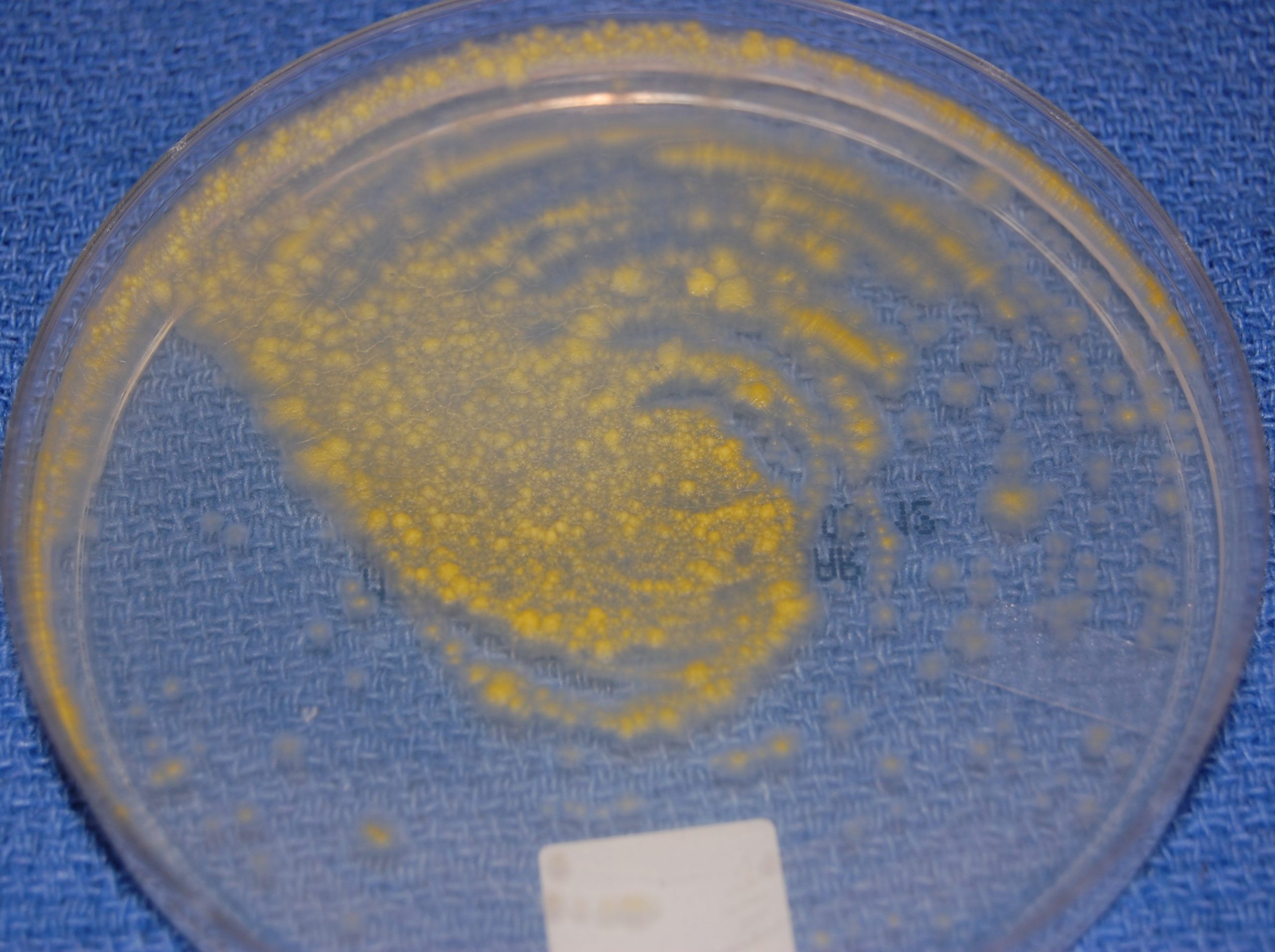
Description: M kansasii  Photograph of 7H10 media  showing yellow pigmentation

Magnification: NA

Specimen Source: Sputum

Date Taken:5.3.2012


Left blue arrow shows a buff colony. With more light exposure, it will produce yellow pigment

If Mycobacterium kansasii is isolated from a culture, it is  considered a pathogen (physician should be notified immediately).

Growth usually takes 7 to 21 days, making it a relatively fast grower among slow-growing mycobacterium

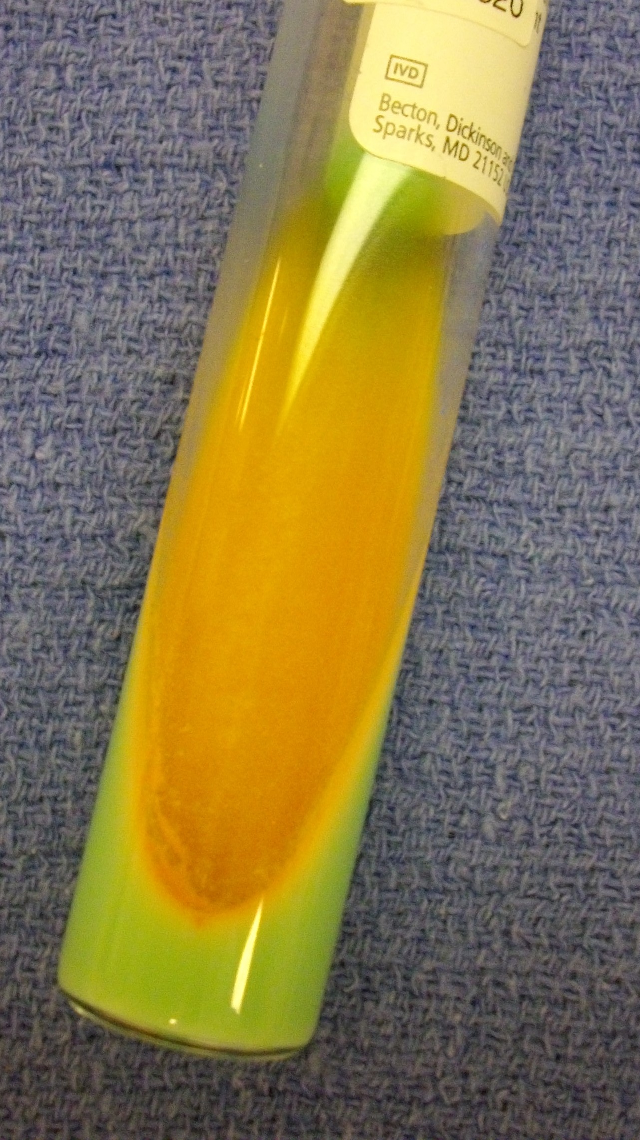
A characteristic feature is photochromogenicity,

                       Colonies are pale in the dark

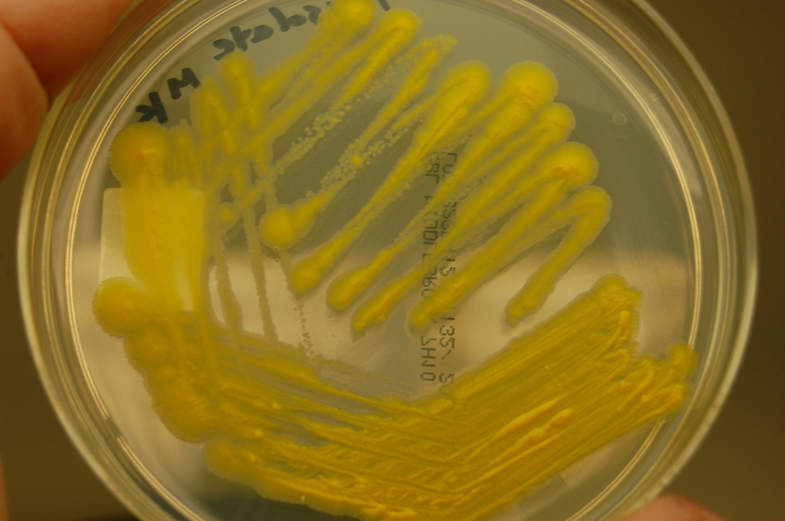
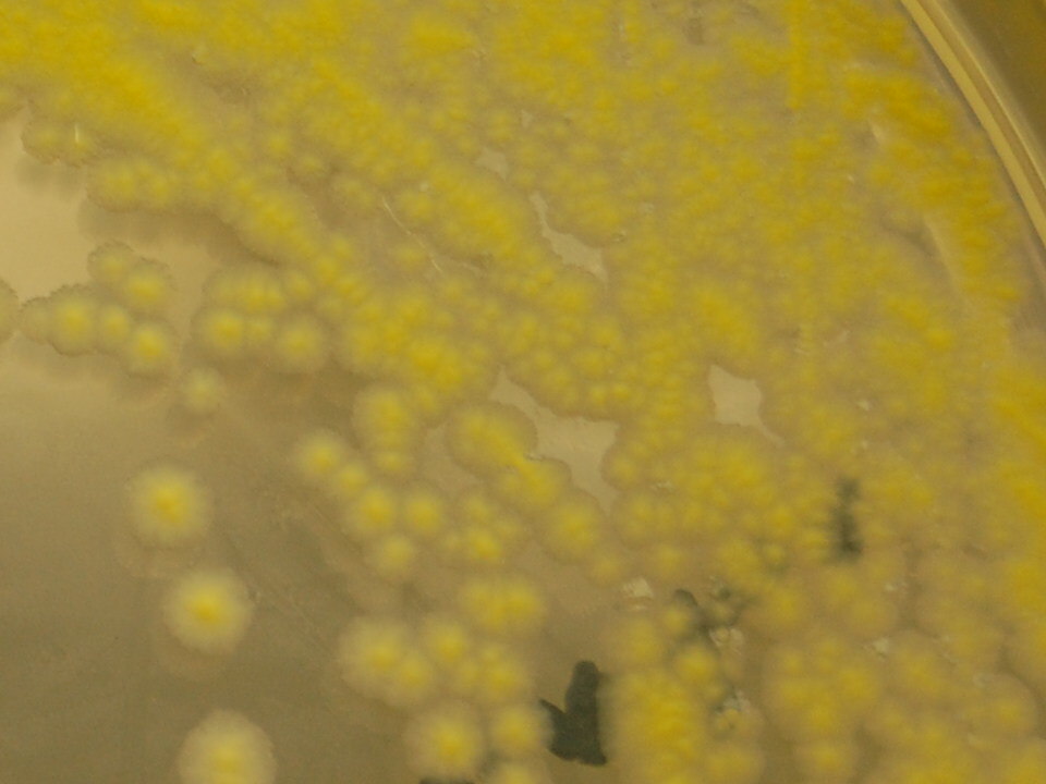
                       After exposure to light, they produce a bright yellow pigment

This property is a classic laboratory clue

Classified as a Runyon Group 1 (photochromogen) mycobacterium

Identification involves a combination of clinical findings, laboratory testing and imaging.

Biochemical identification are now less commonly relied upon because molecular methods are faster and more accurate. The present testing for identification is PCR, DNA sequencing and MALDI-TOF.

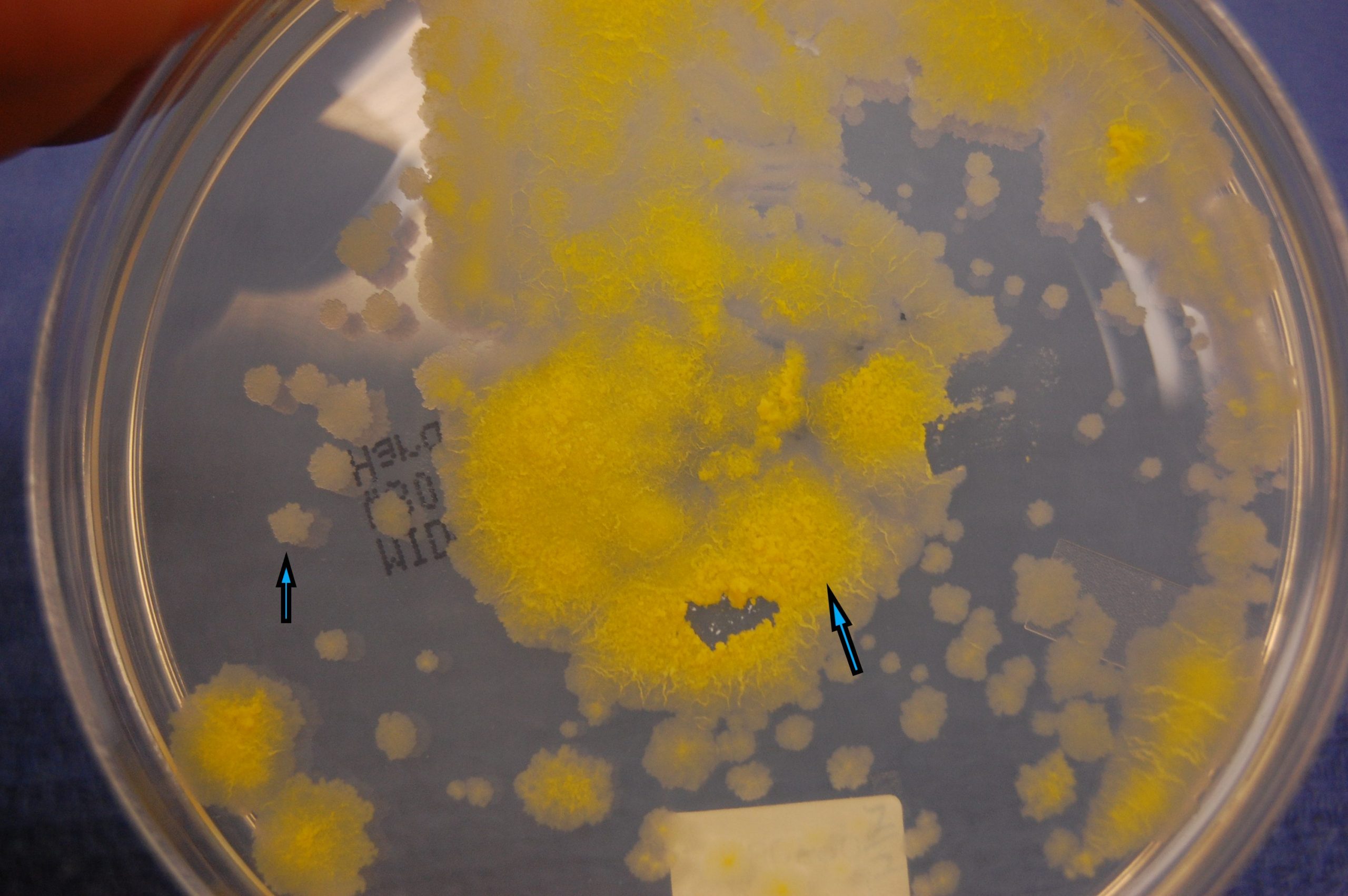
The photo gallery below shows photographs of 7H10 agar and LJ slants of Mycobacterium kansasii