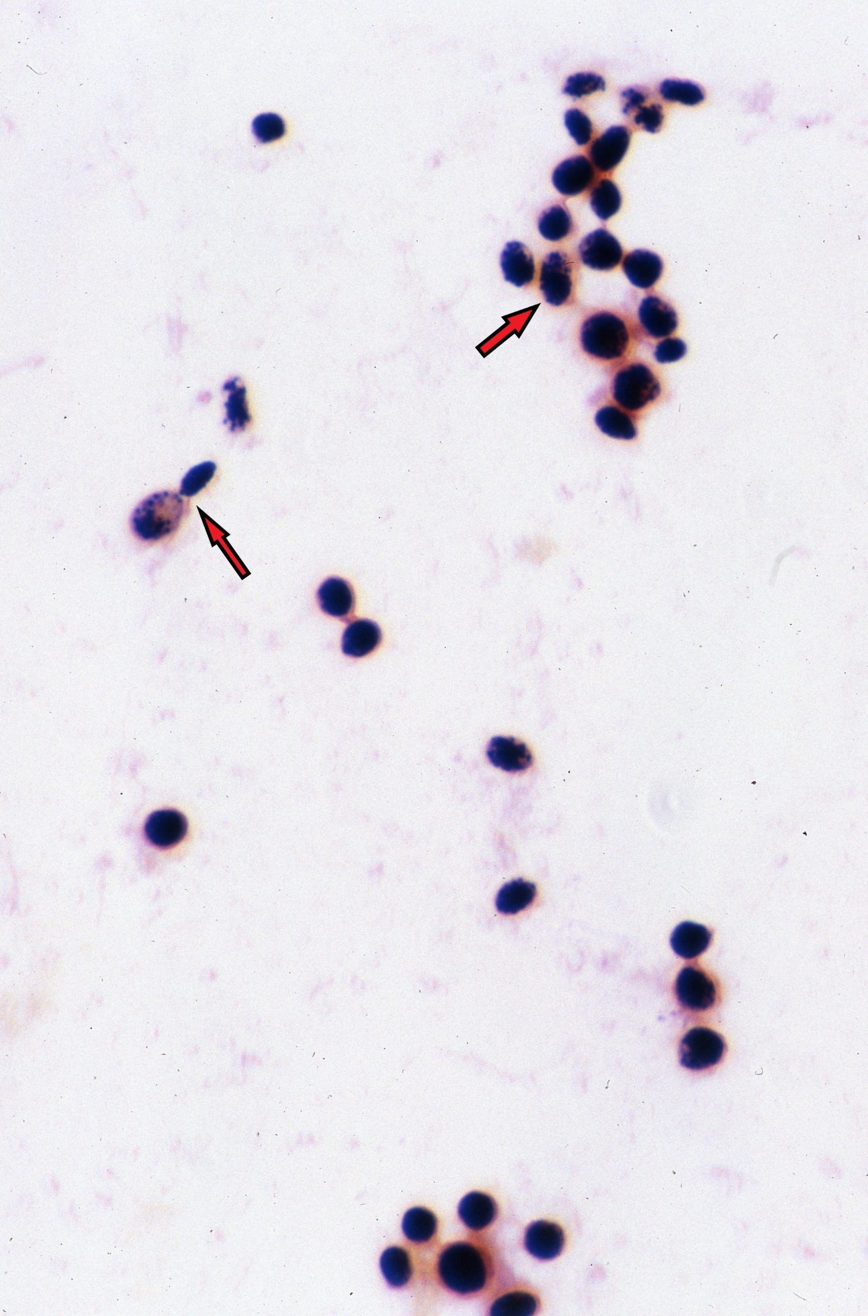
India Ink
-

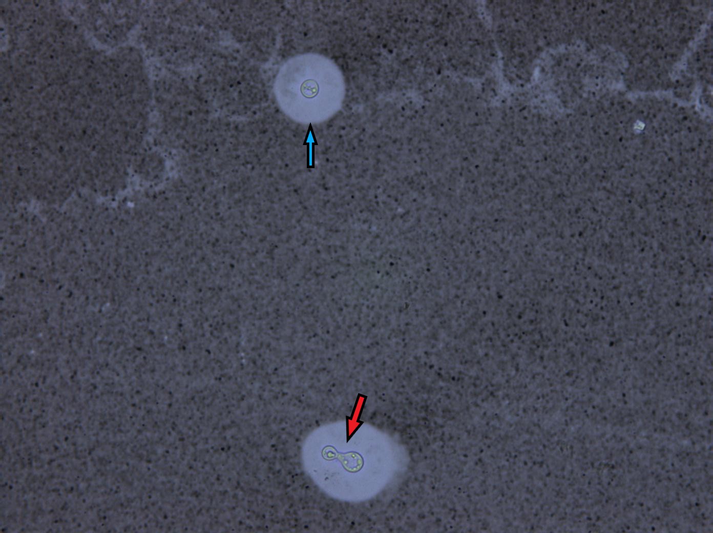
- MAG 40X Blue arrow pointing to the capsule. Red arrow pointing to the yeast cell
-
- MAG 40X note the budding yeast. Diagnostic for C. neoformans
-
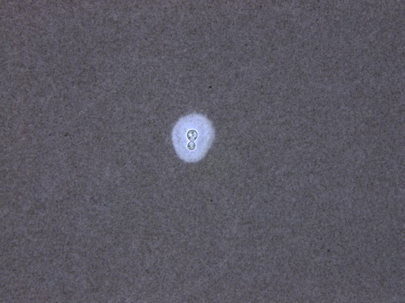
- MAG 40X
-
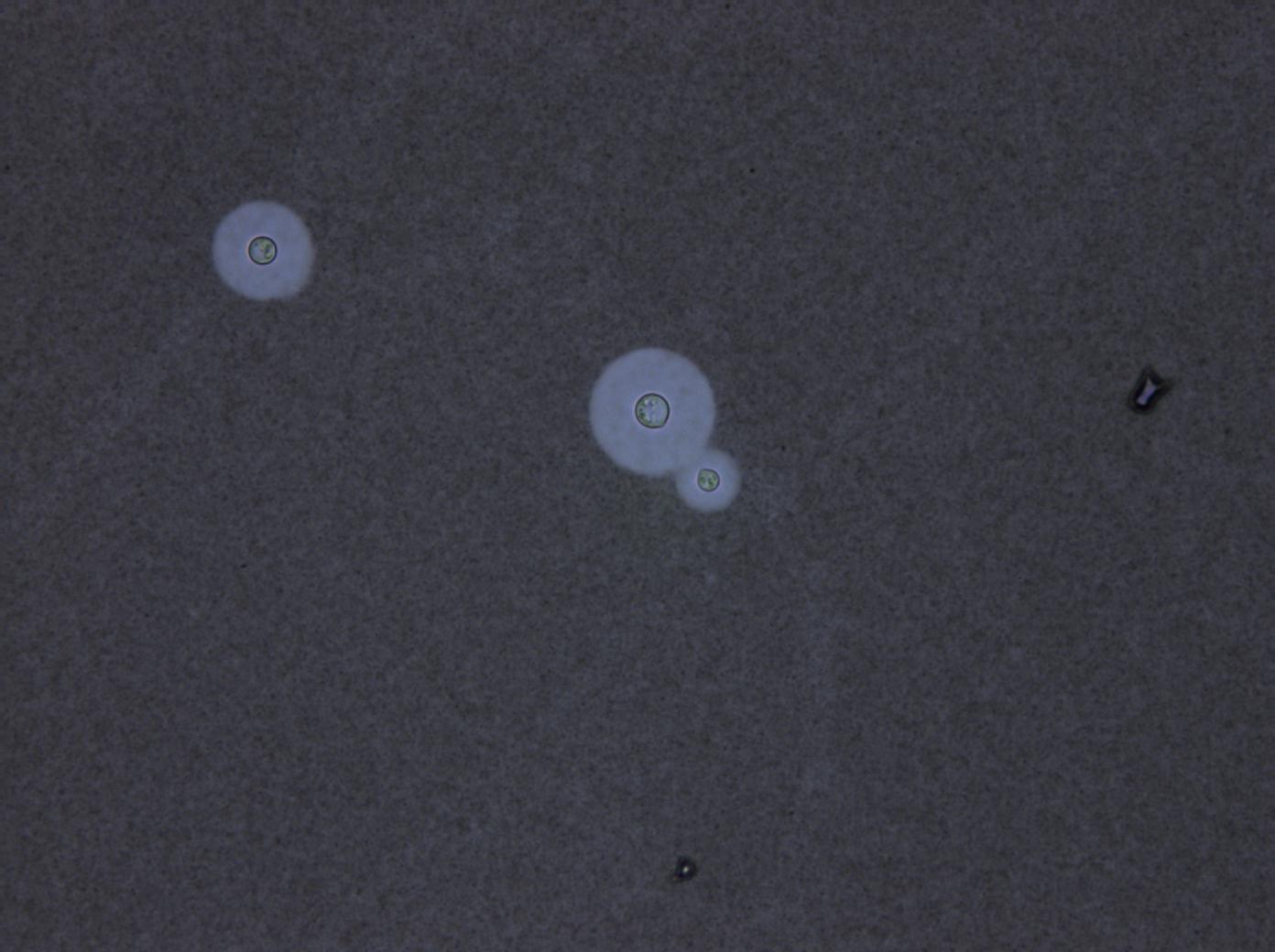
- MAG 40X
-
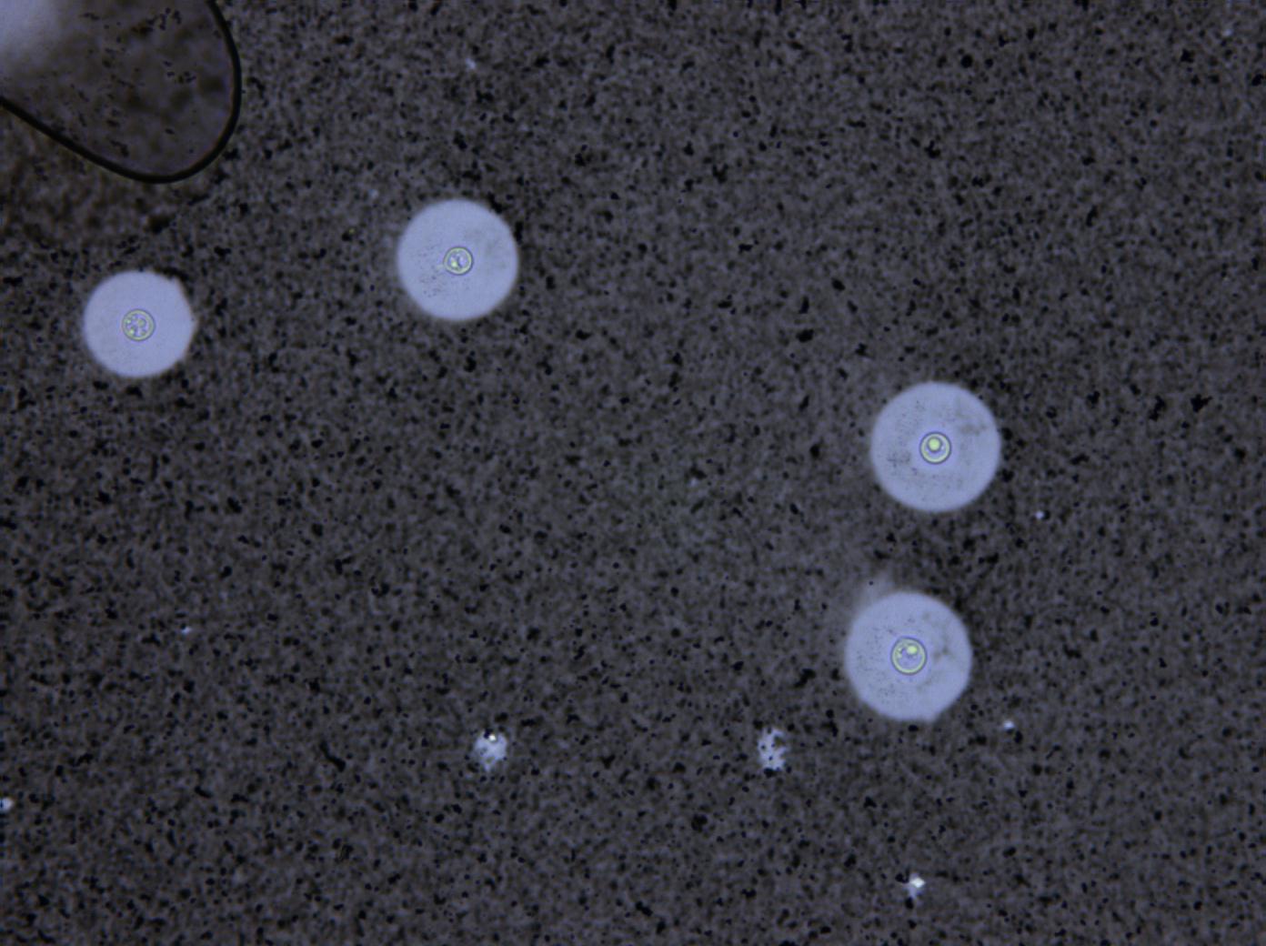
- MAG 10X
-
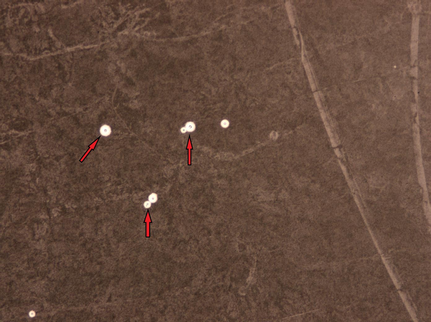
- MAG 10X
-
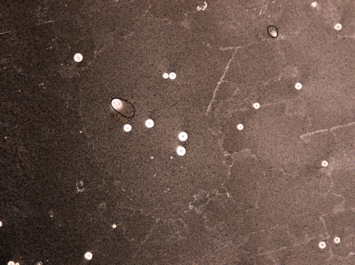
- MAG 10X Red arrow pointing to a budding yeast
-
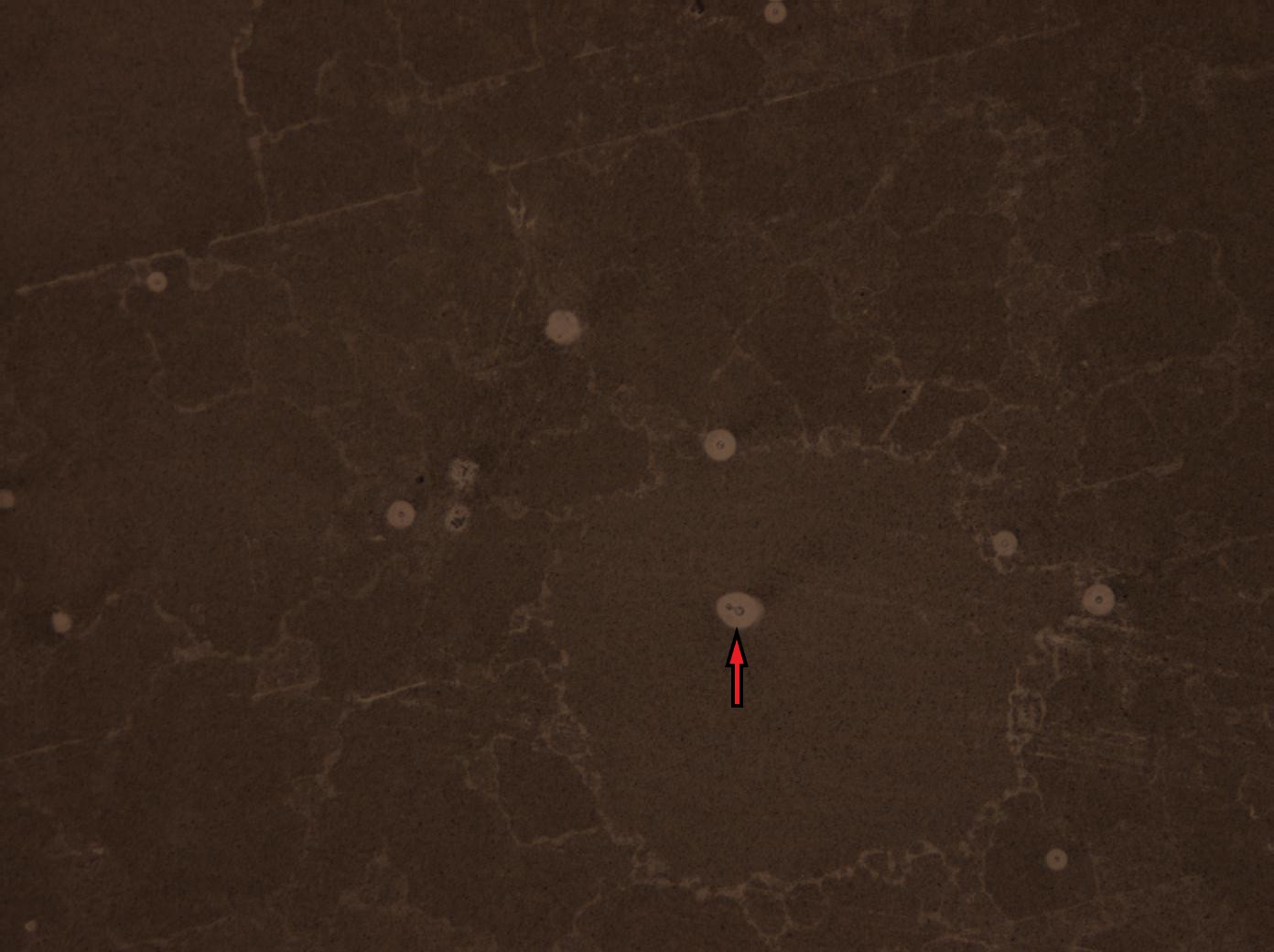
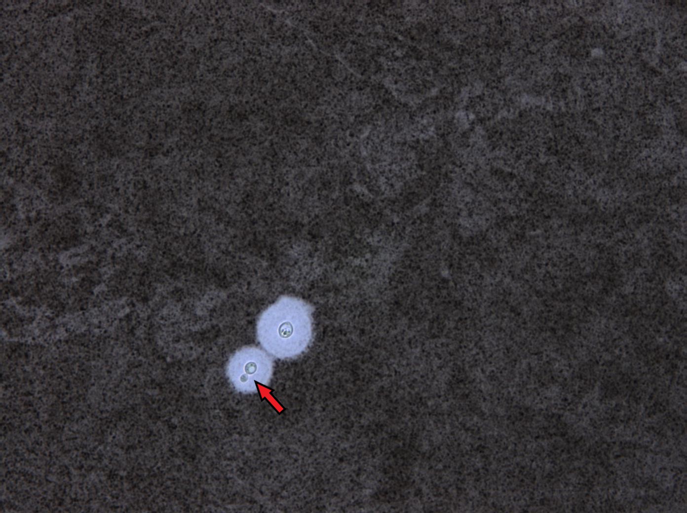
- MAG 40X Red arrow pointing to budding yeast
-
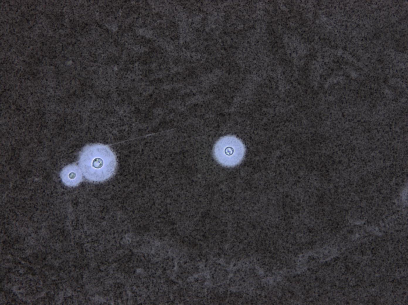
- MAG 40X
-
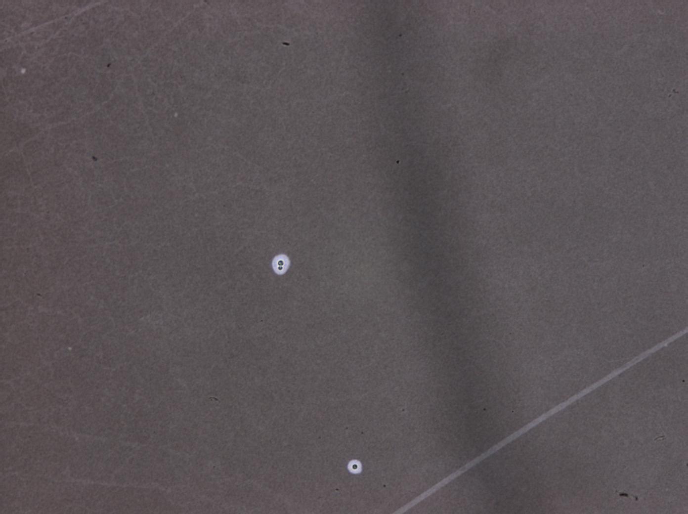
- MAG 10X
-
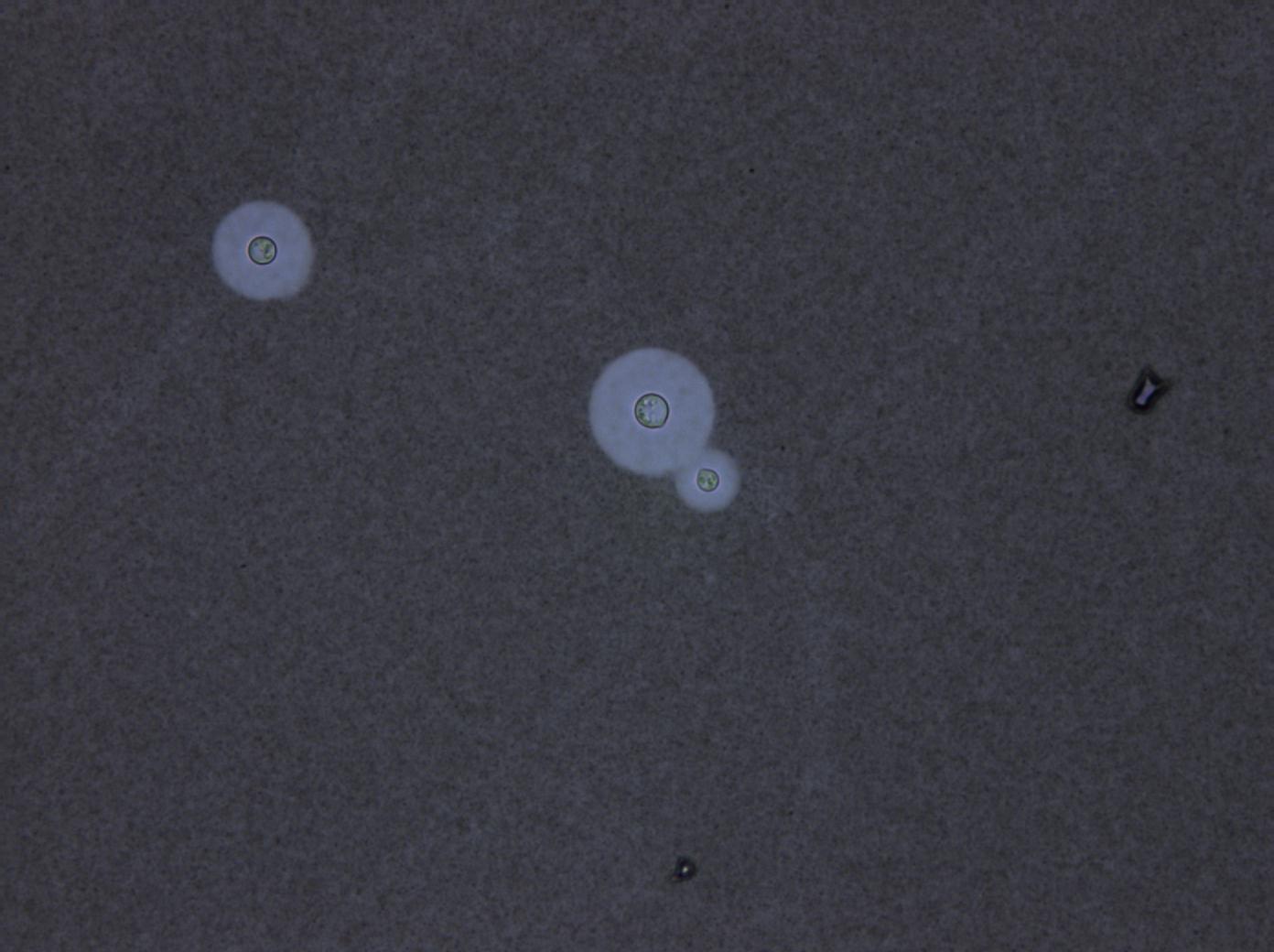
- MAG 40X
-
- MAG 10X
-
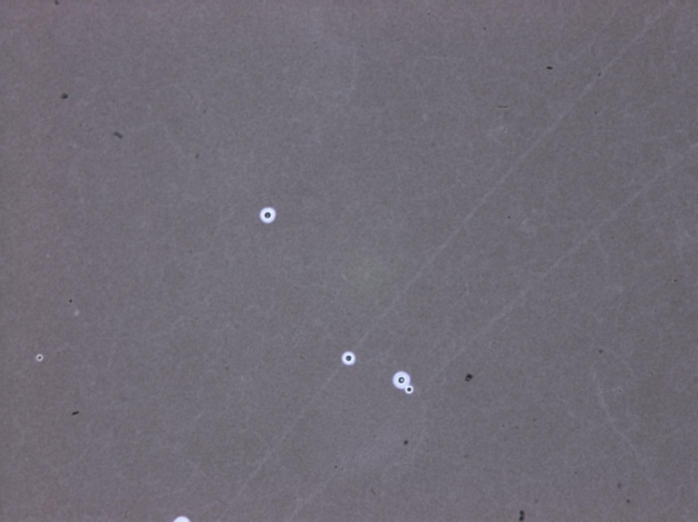
- MAG 40X
-
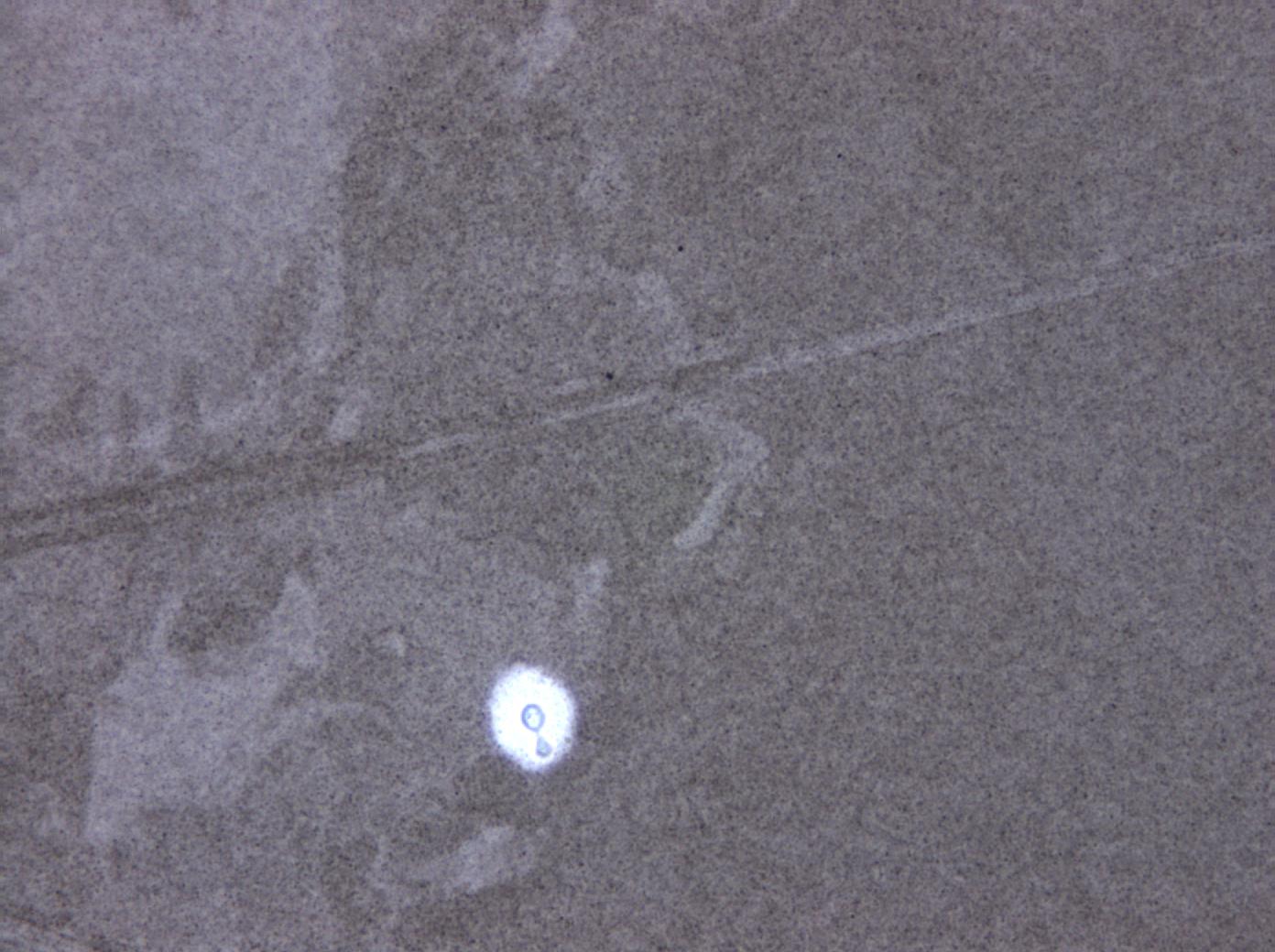
- MAG 10X
-
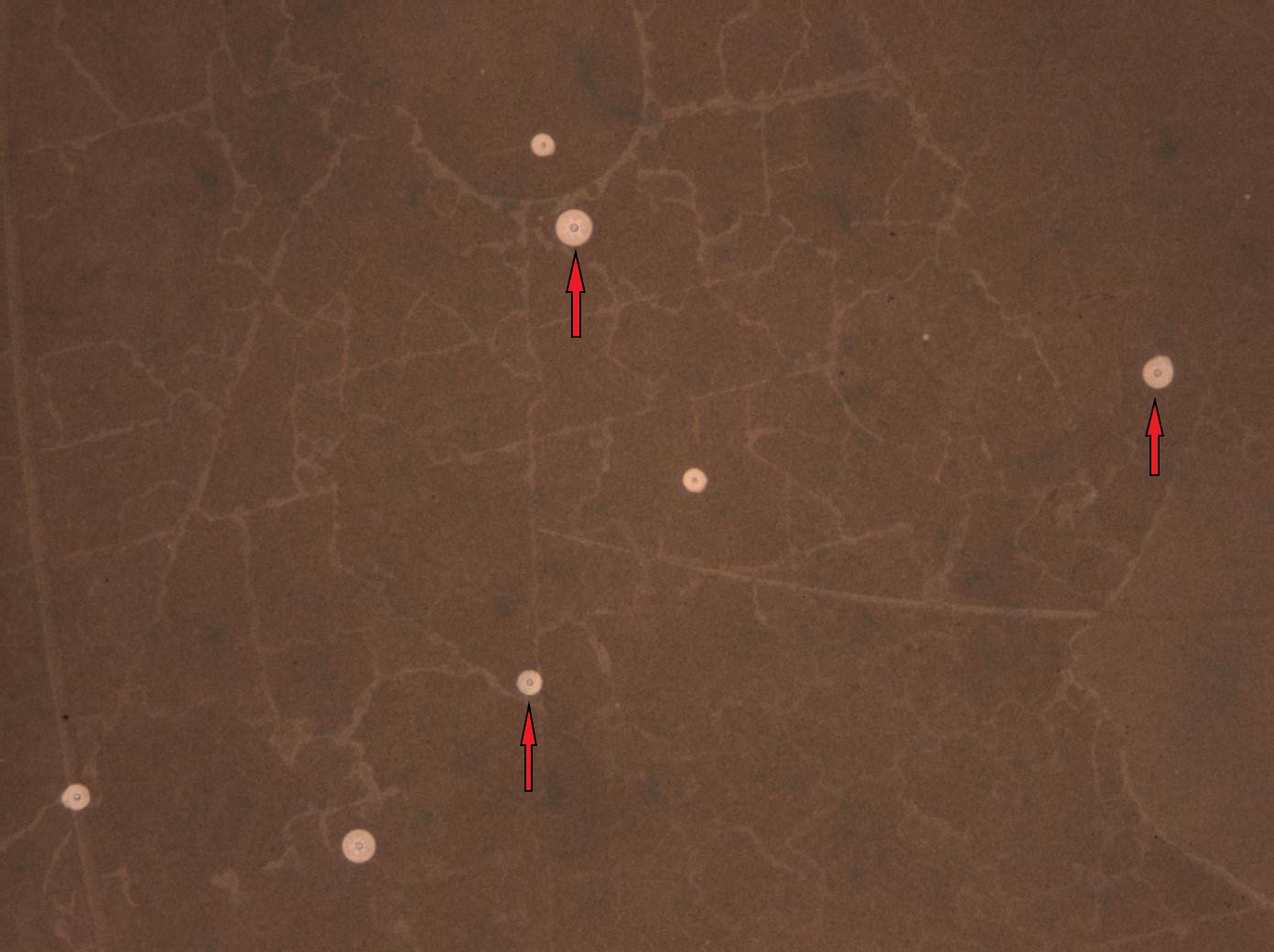
- MAG 100X
-
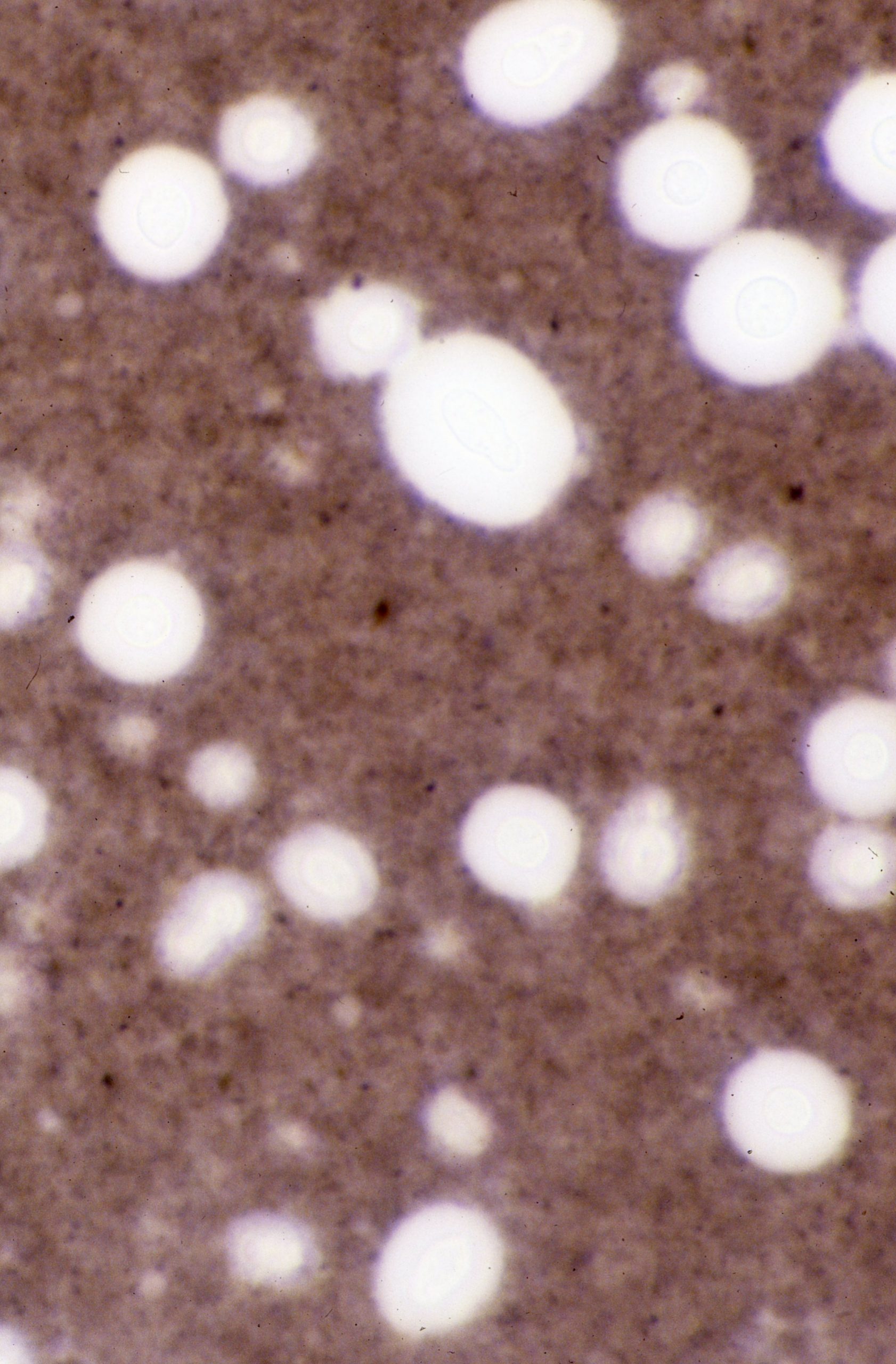
- MAG 100X
-
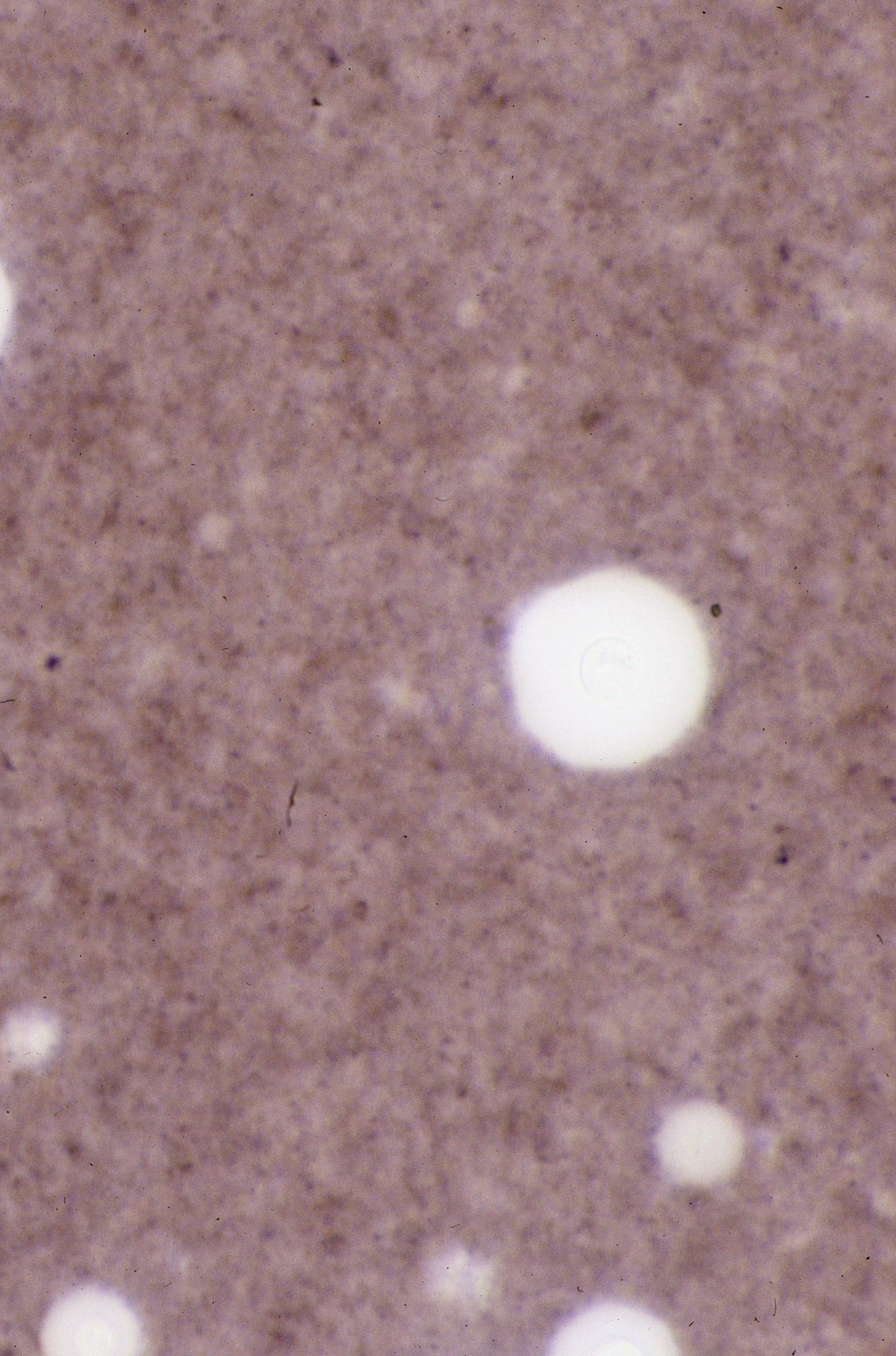
- MAG 40X Note how many yeast cells are in this one field of view
-
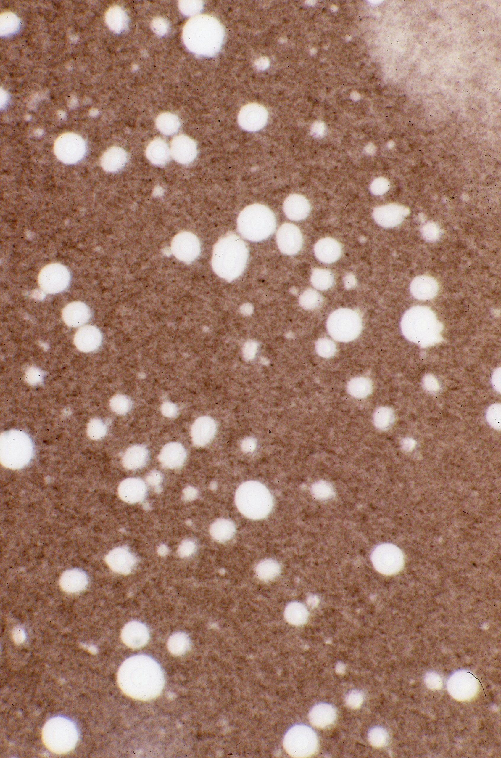
- MAG 10X
-
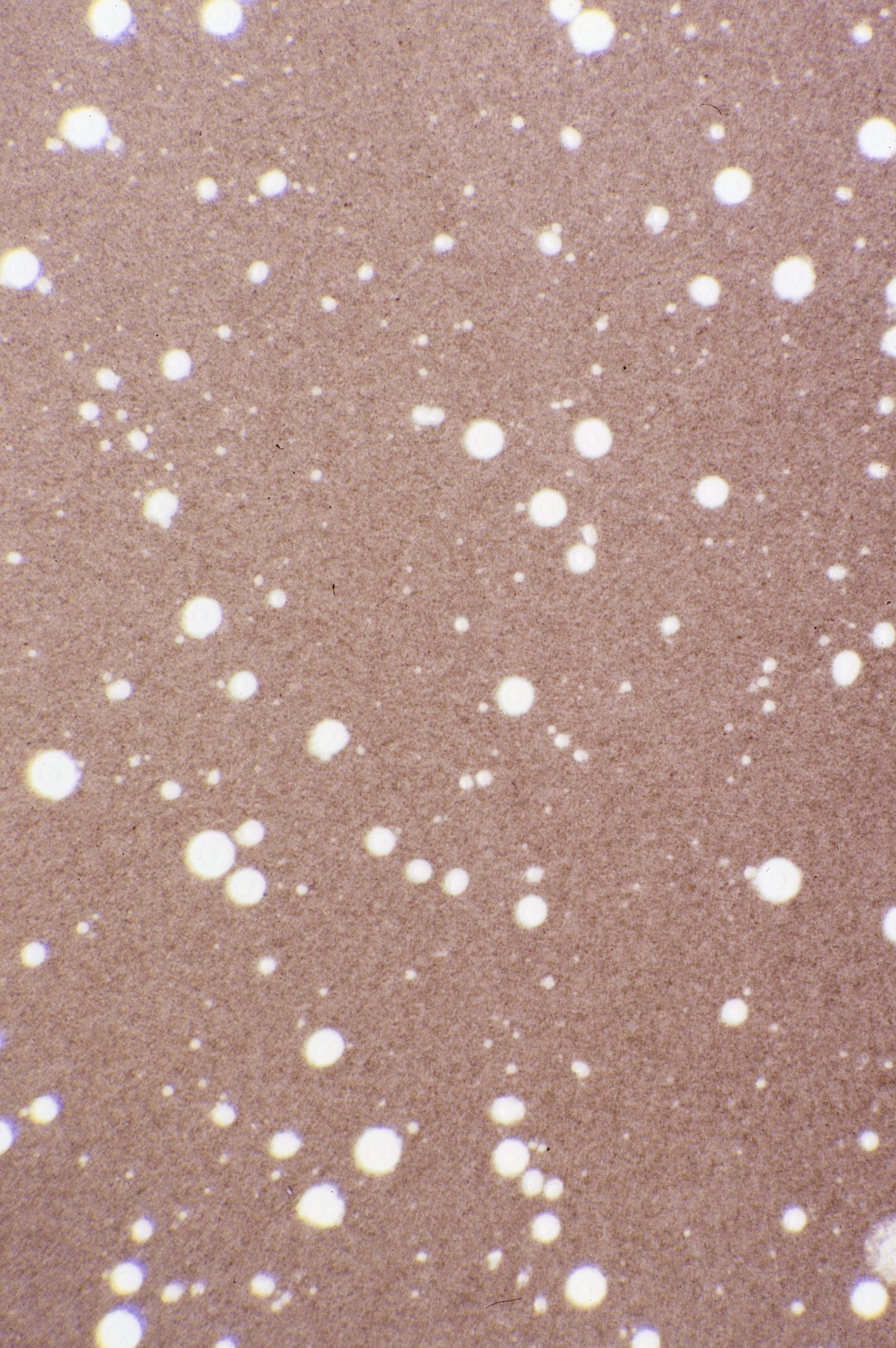
- MAG 40X
-
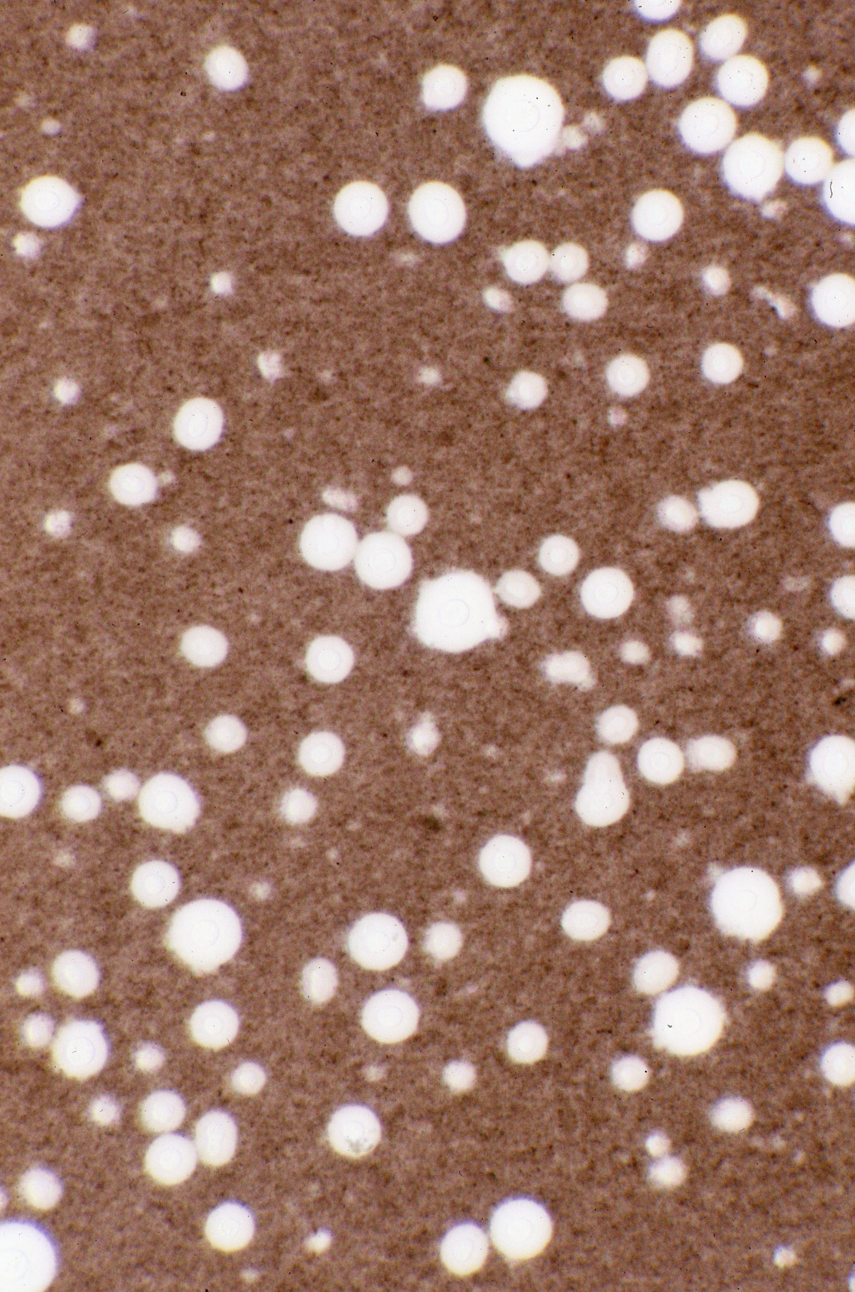
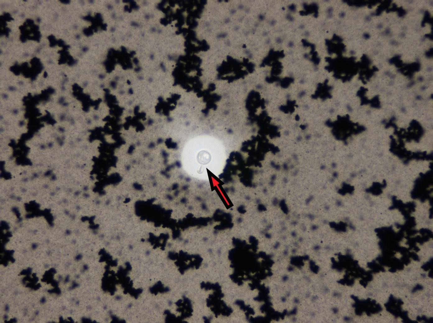
- MAG 40X Red arrow pointing to budding yeast, positive to C. neoformans
-
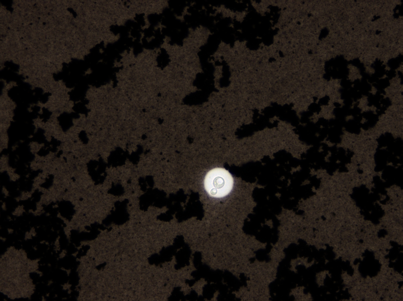
- MAG 40X Note budding yeast
-
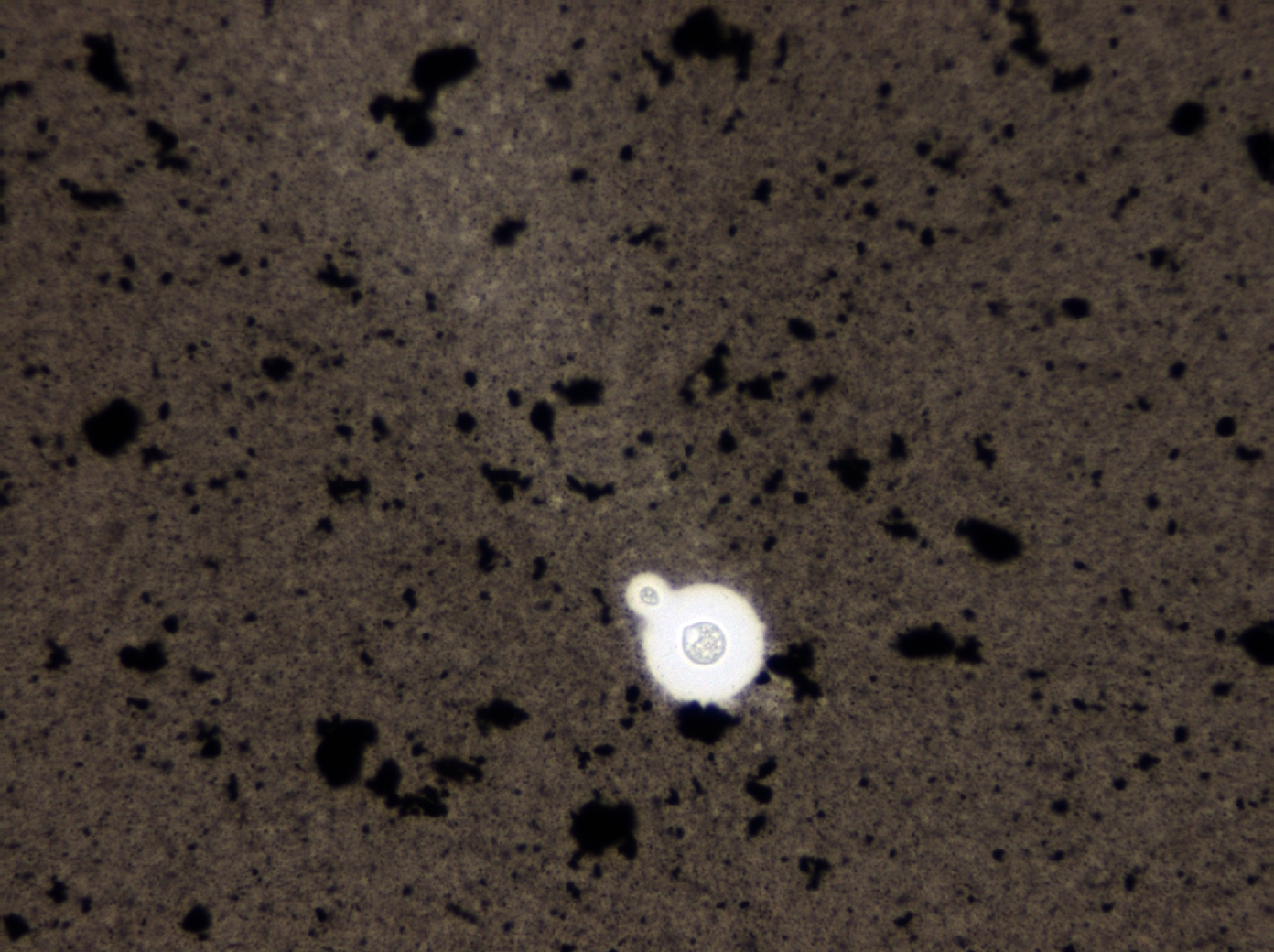
- MAG 40X
-
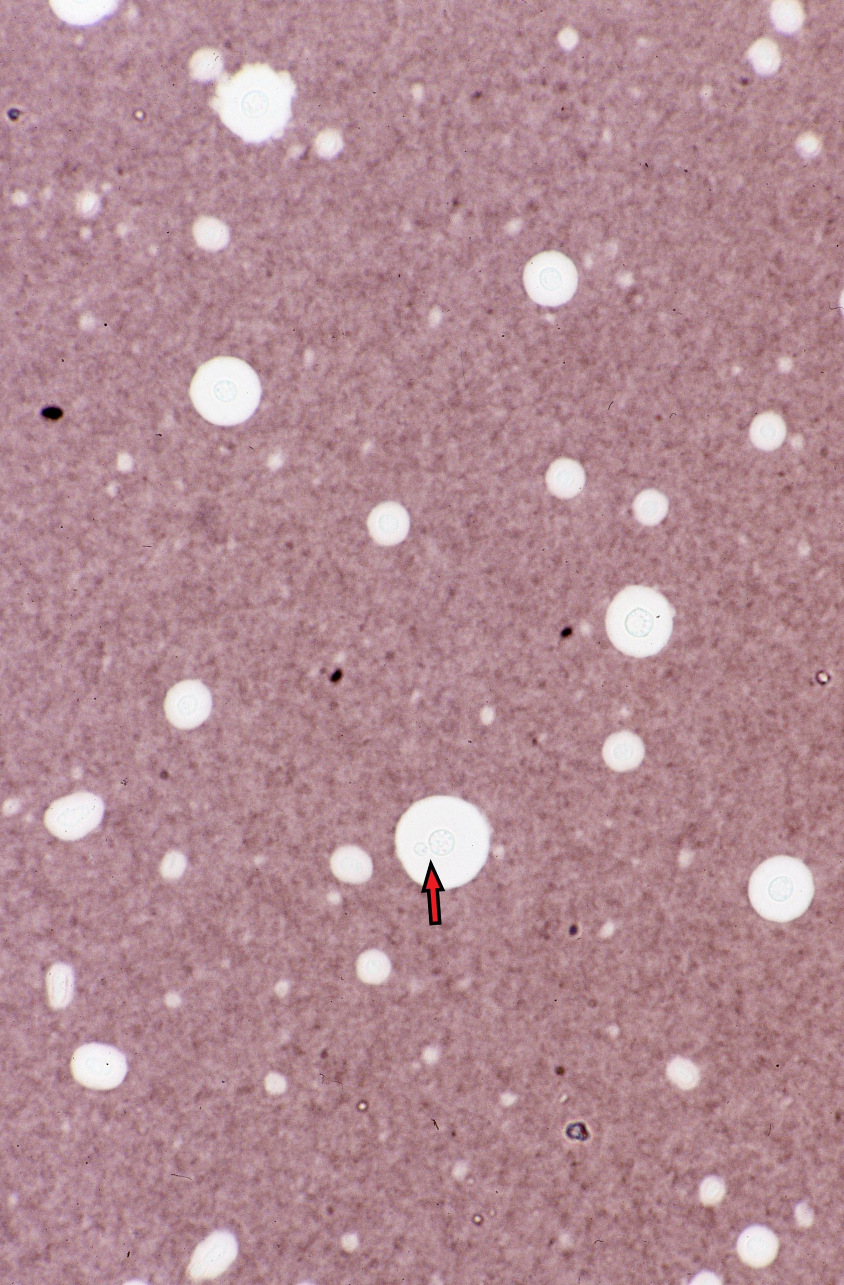
- MAG 40X Red arrow pointing to yeast cell
-
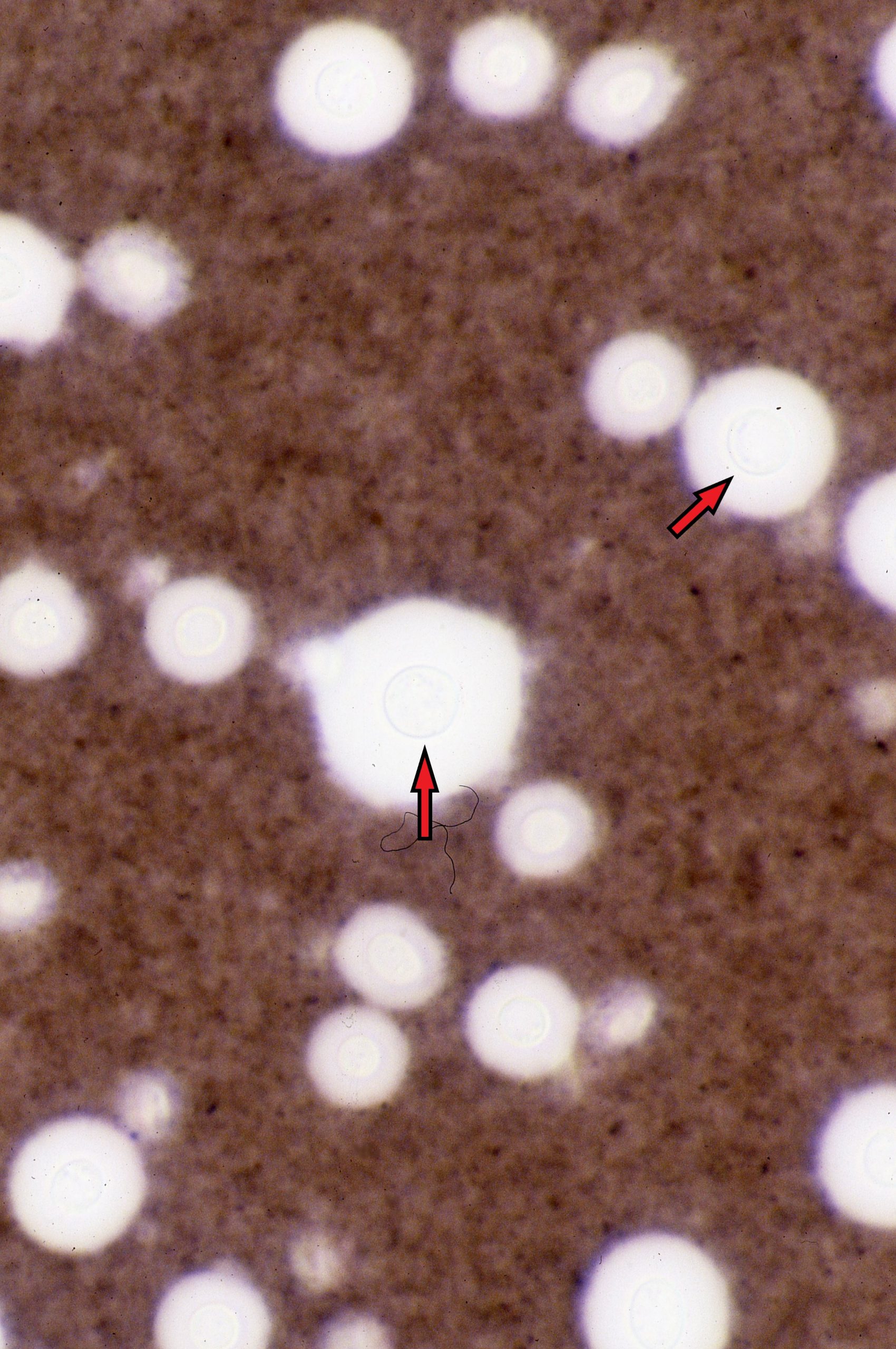
- MAG 100X Red arrows pointing to the yeast cells within the capsule. Normally you do not view a India ink prep at 100x magnification
-
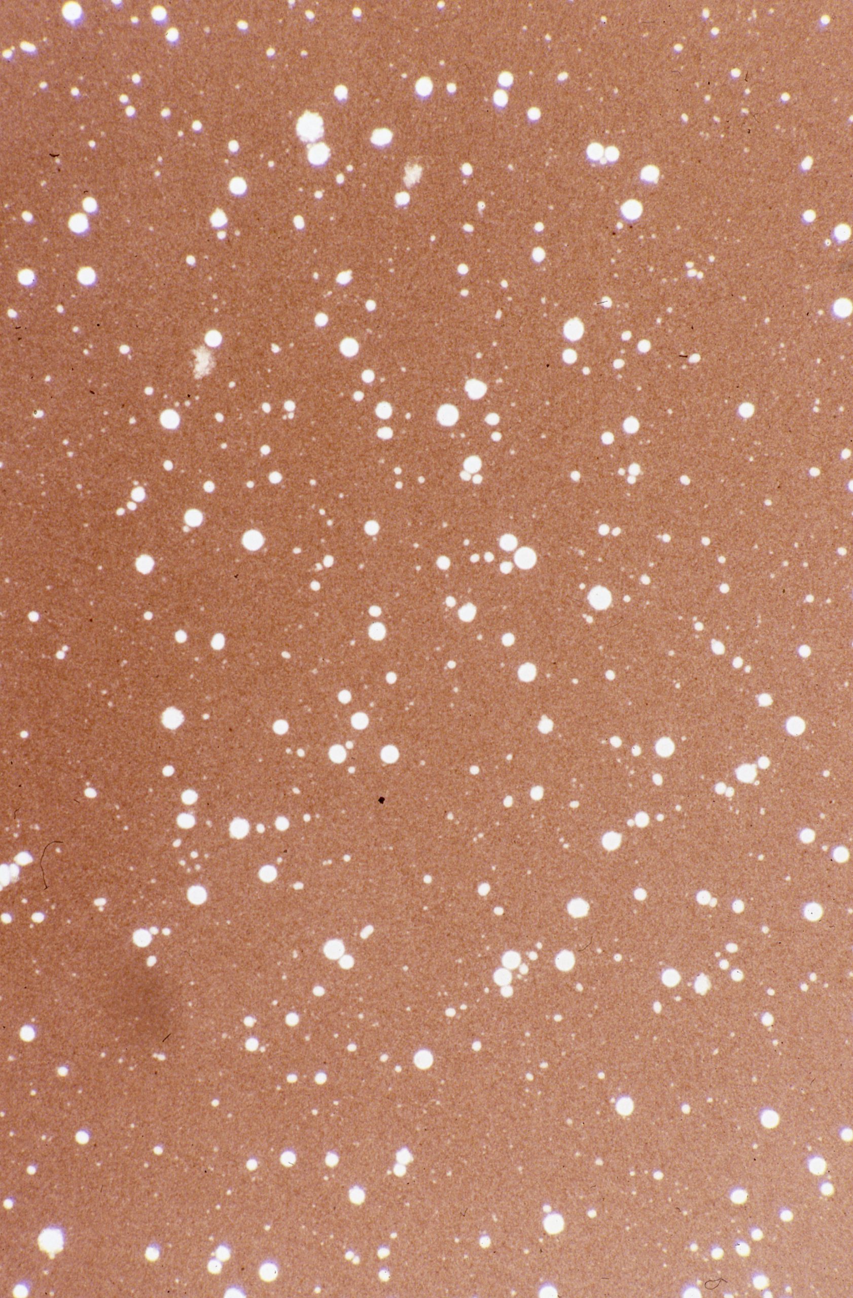
- MAG 10X
-
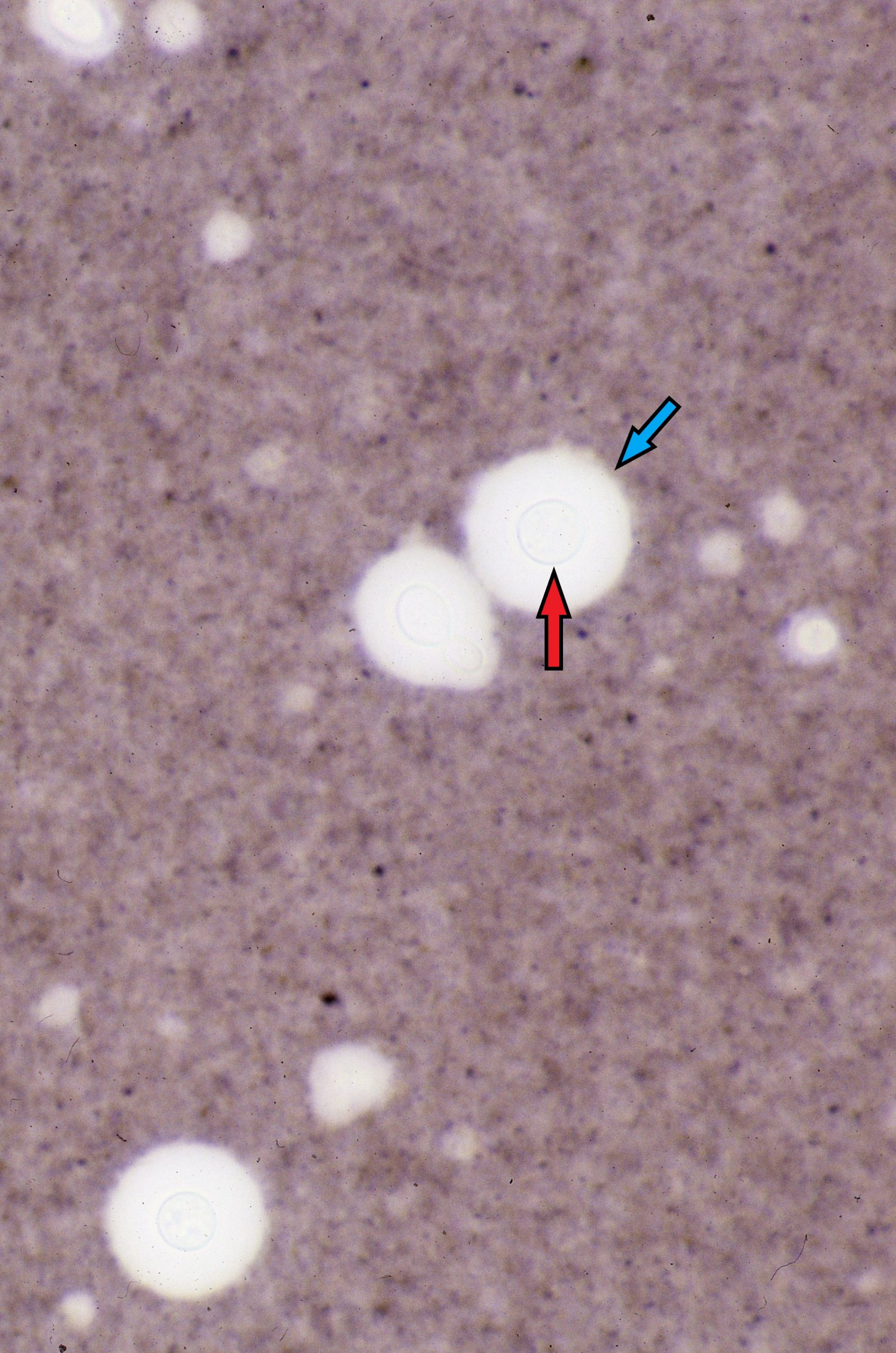
- MAG 100X Red arrow pointing to the the yeast cell. Blue arrow pointing the the polysaccharide capsule
-
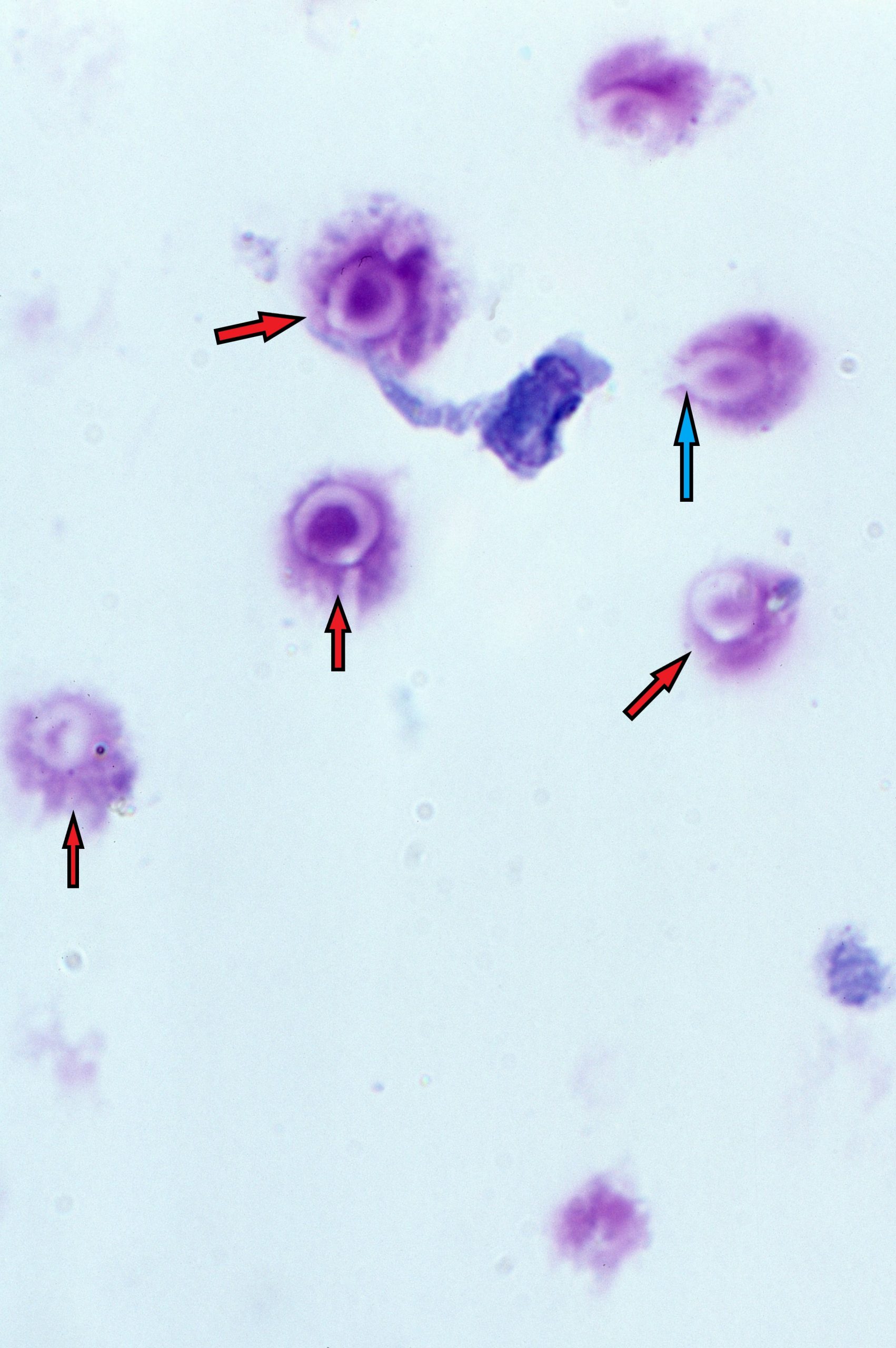
- MAG 100X Red arrows pointing to yeast that are not staining very well due to the capsule. Blue arrow points to budding yeast cell, look closely and see the clearing of the bud
-
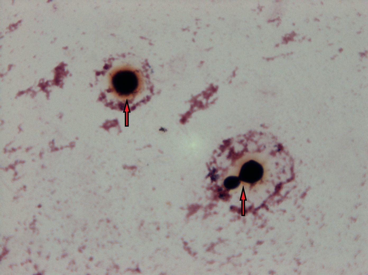
- MAG 100X Bottom arrow points to a budding yeast cell diagnostic of C. neoformans
-
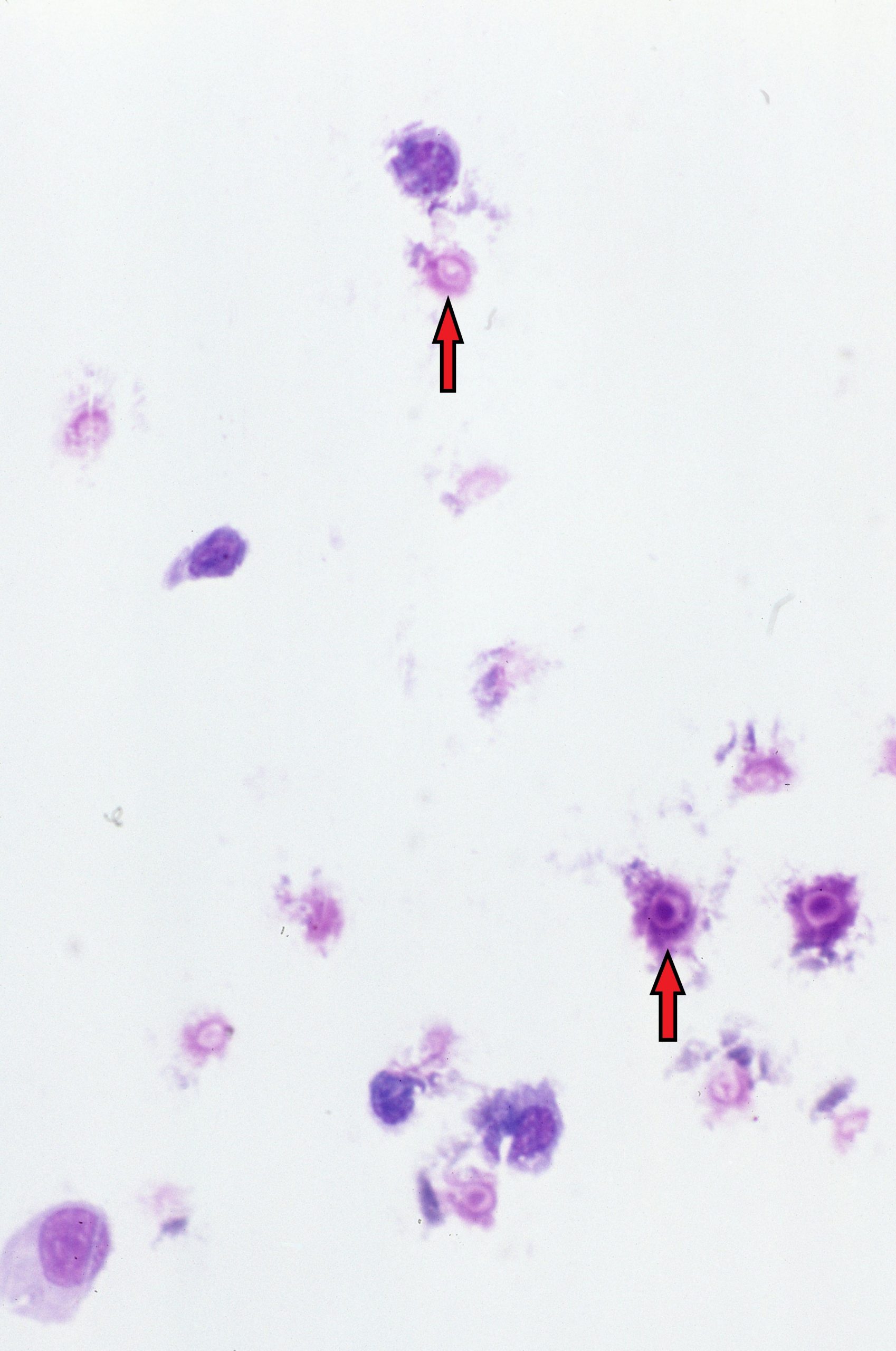
- MAG 40X Red arrows pointing to yeast cells
-
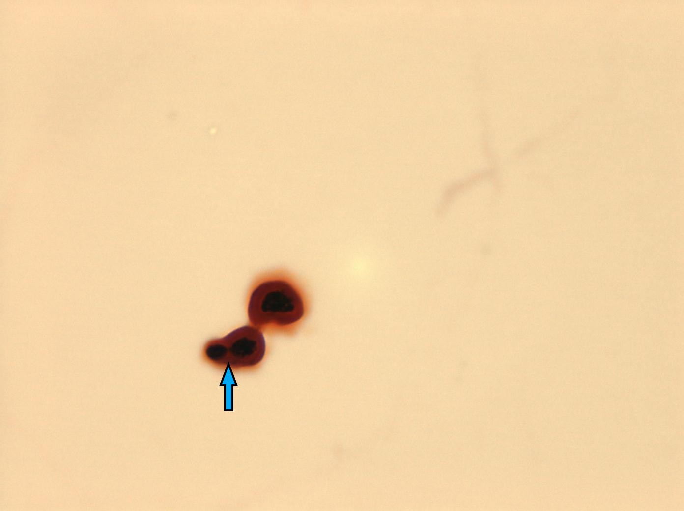
- MAG 100X Blue arrow pointing to a budding yeast cell. Note how the yeast cell is staining blue and note the reddish stained capsule
-
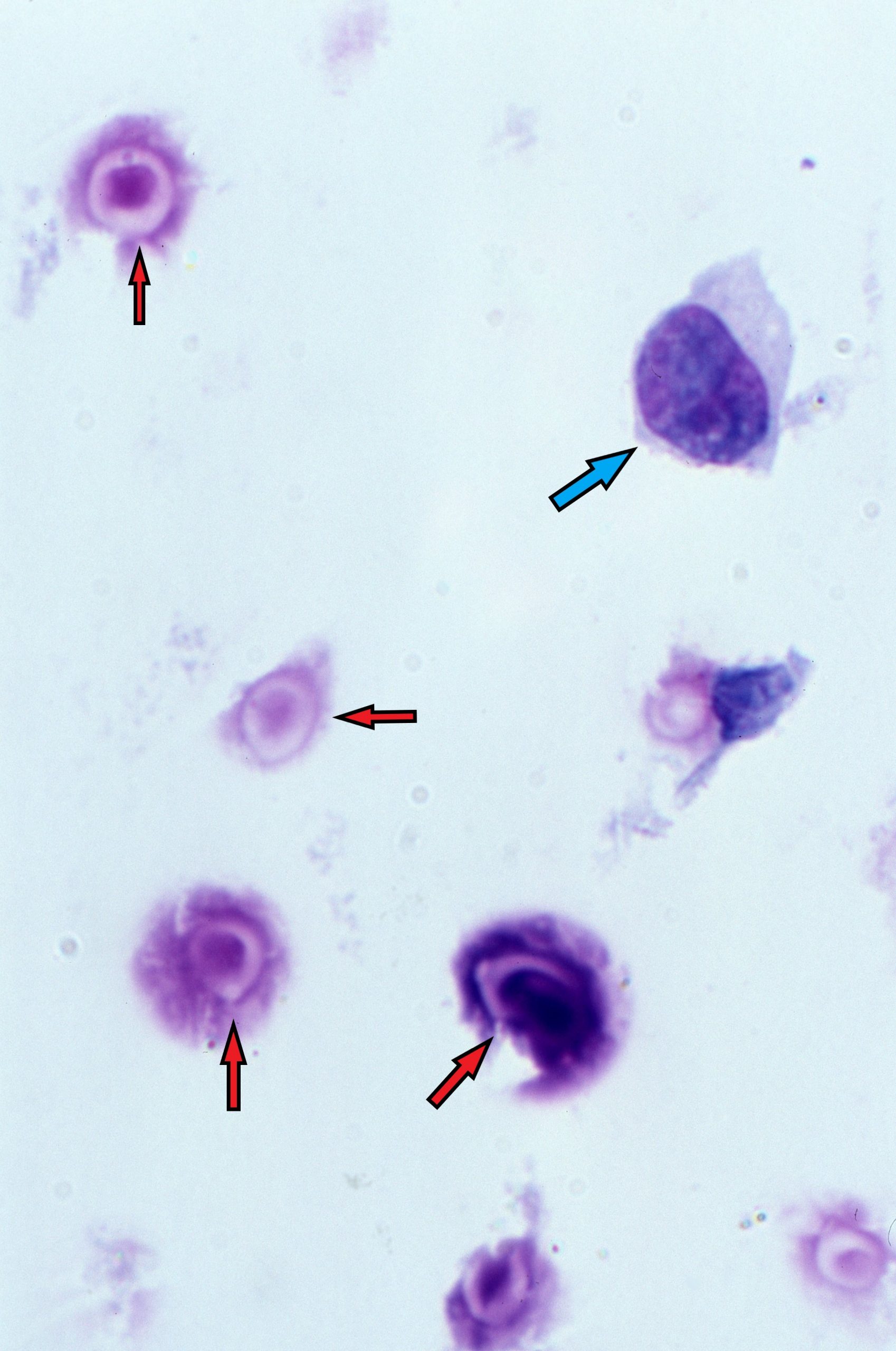
- MAG 100X Red arrows pointing to the poorly stained yeast cells. Blue arrow points to a lymphocyte
-
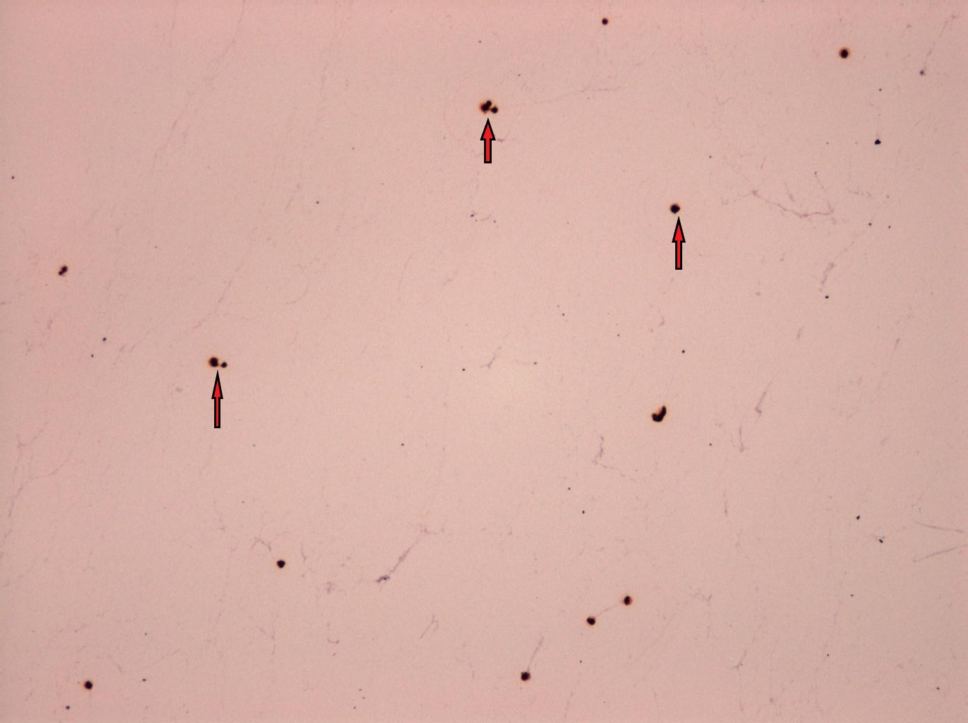
- MAG 10X Red arrows pointing to yeast cells
-
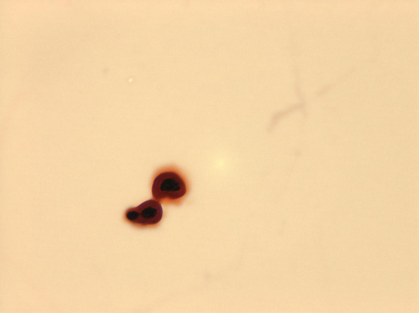
- MAG 100X Slide shows the Gram positive yeast cells
-
- MAG 100X

-
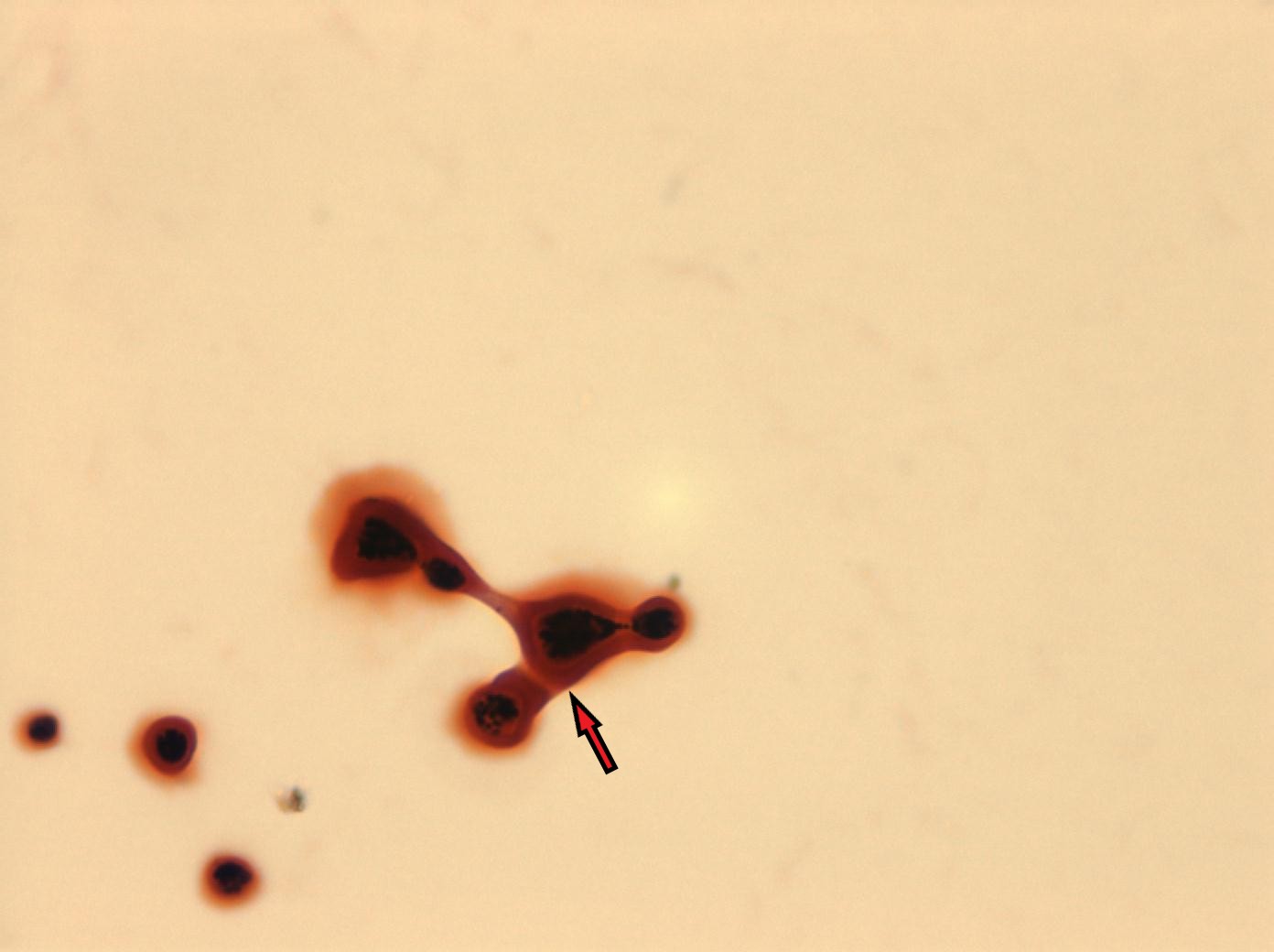
- MAG 100X
-
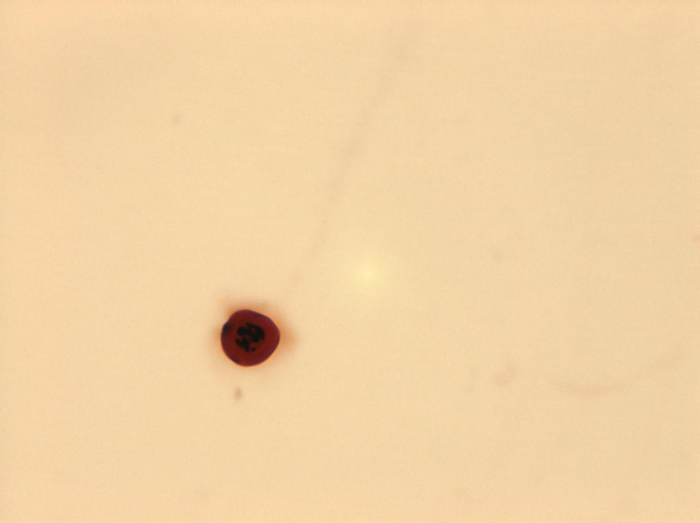
- MAG 100X
-
- MAG 100X

-
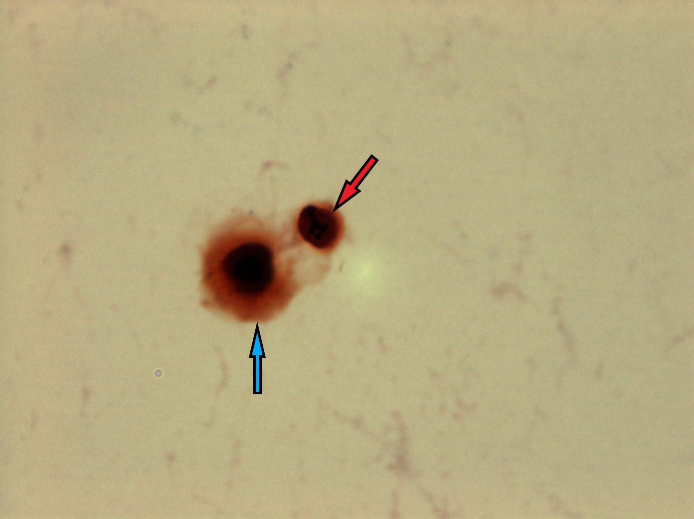
- MAG 100X Red arrow points to the yeast cell. Blue arrow points to the capsule
-
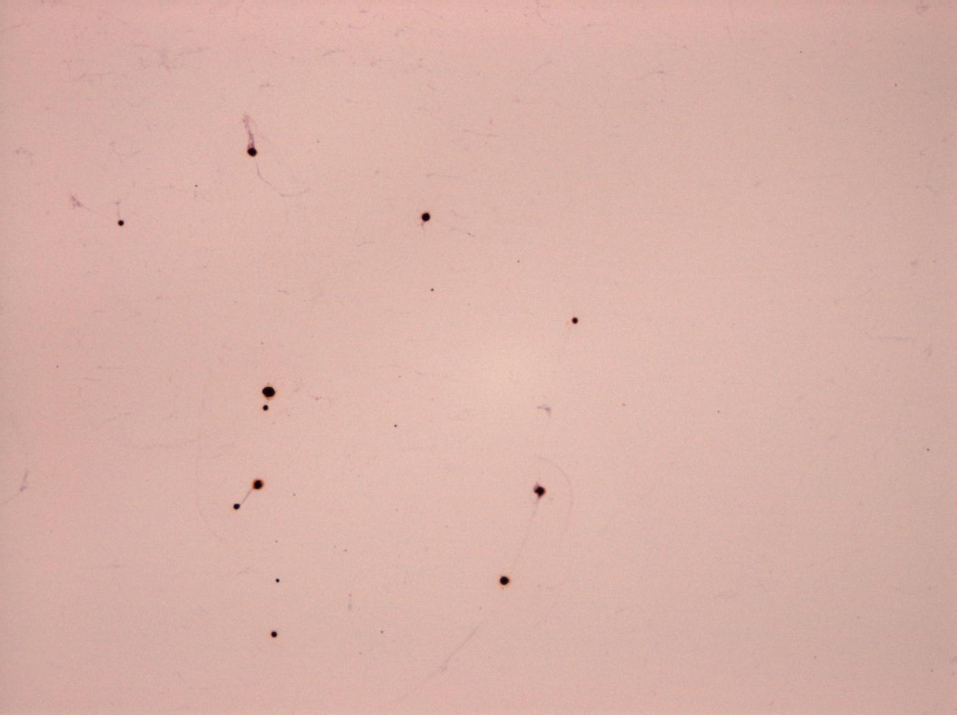
- MAG10X
-
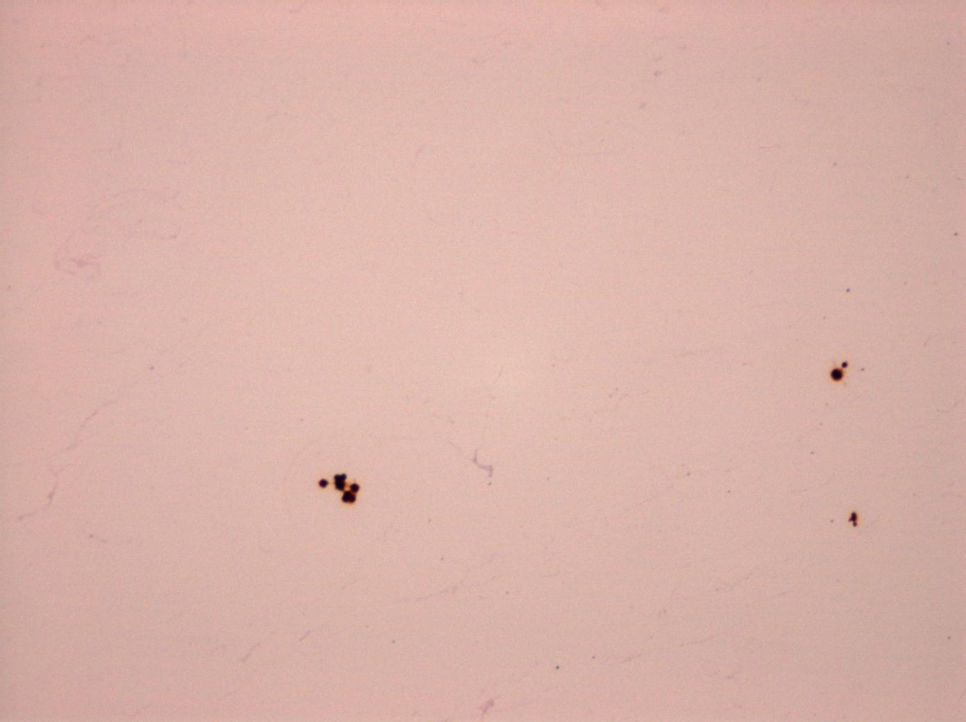
- MAG 10X
-
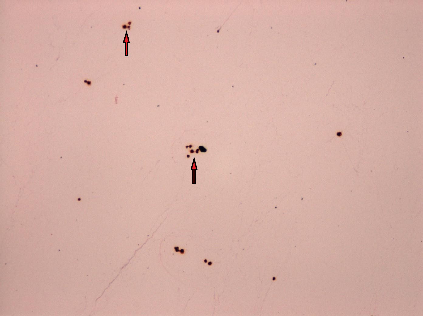
- MAG 10X Red arrows pointing to yeast cells
-
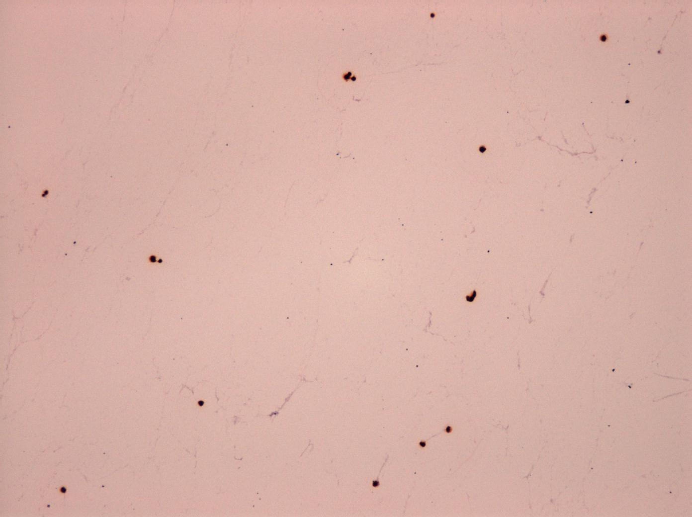
- MAG 10X
-
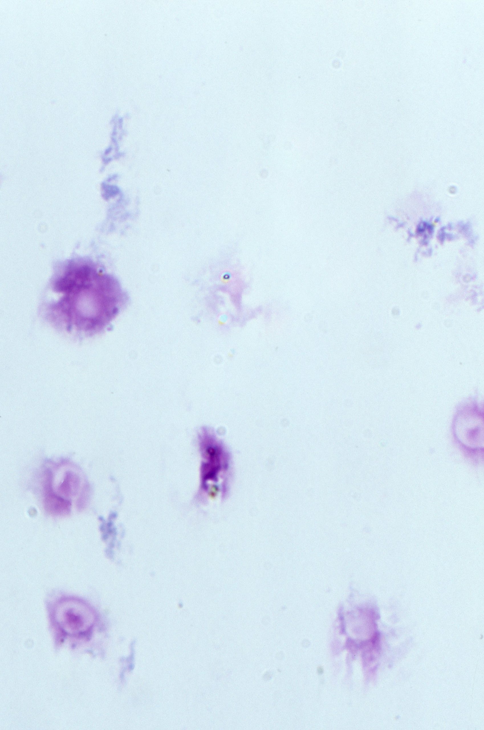
- MAG 100X Slide could be misread as artifacts due to the poorly staining yeast cells
-
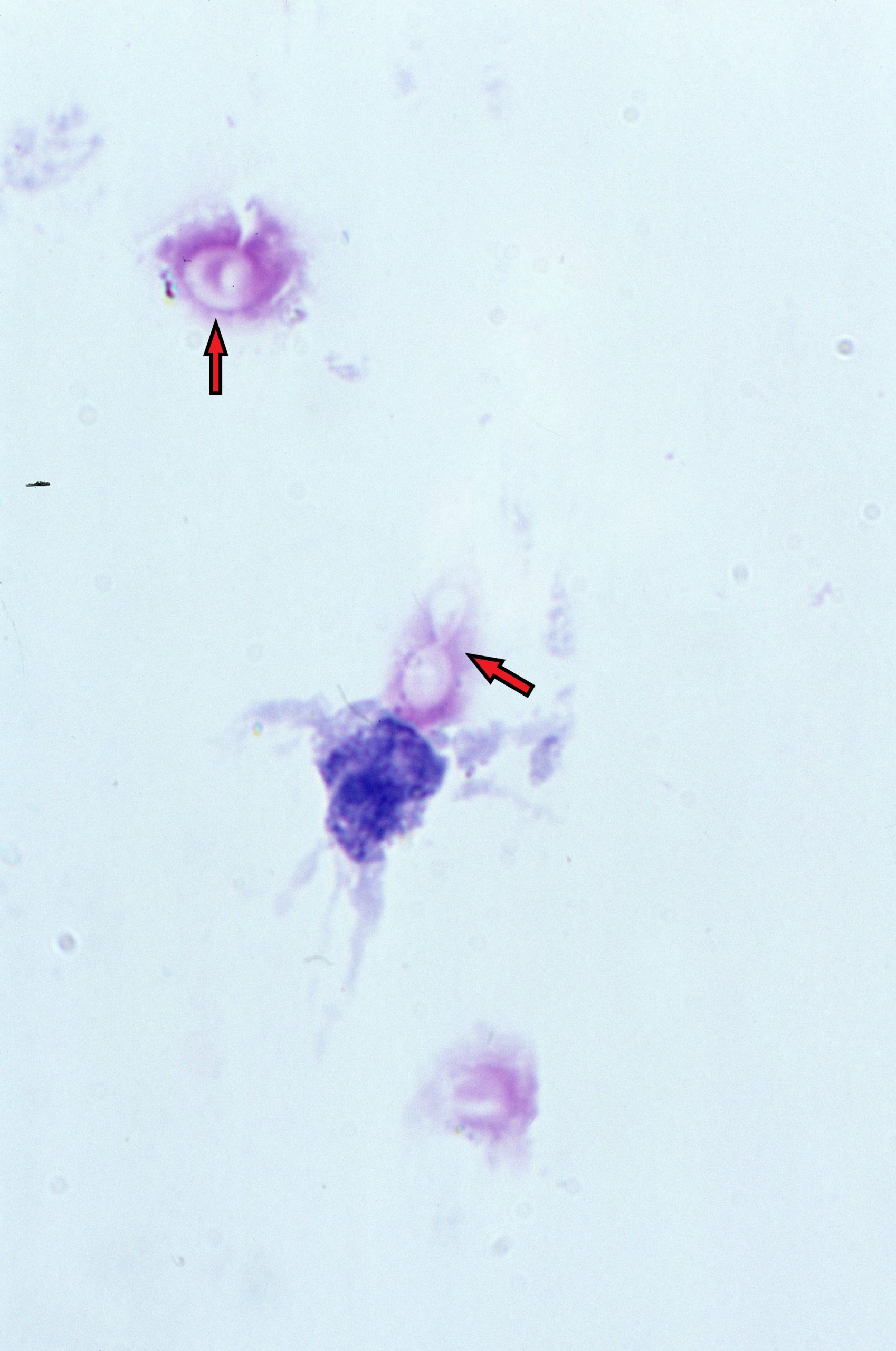
- MAG 100X Red arrows points to yeast cells. Note the bottom arrow shows a poorly stained budding yeast cell
-
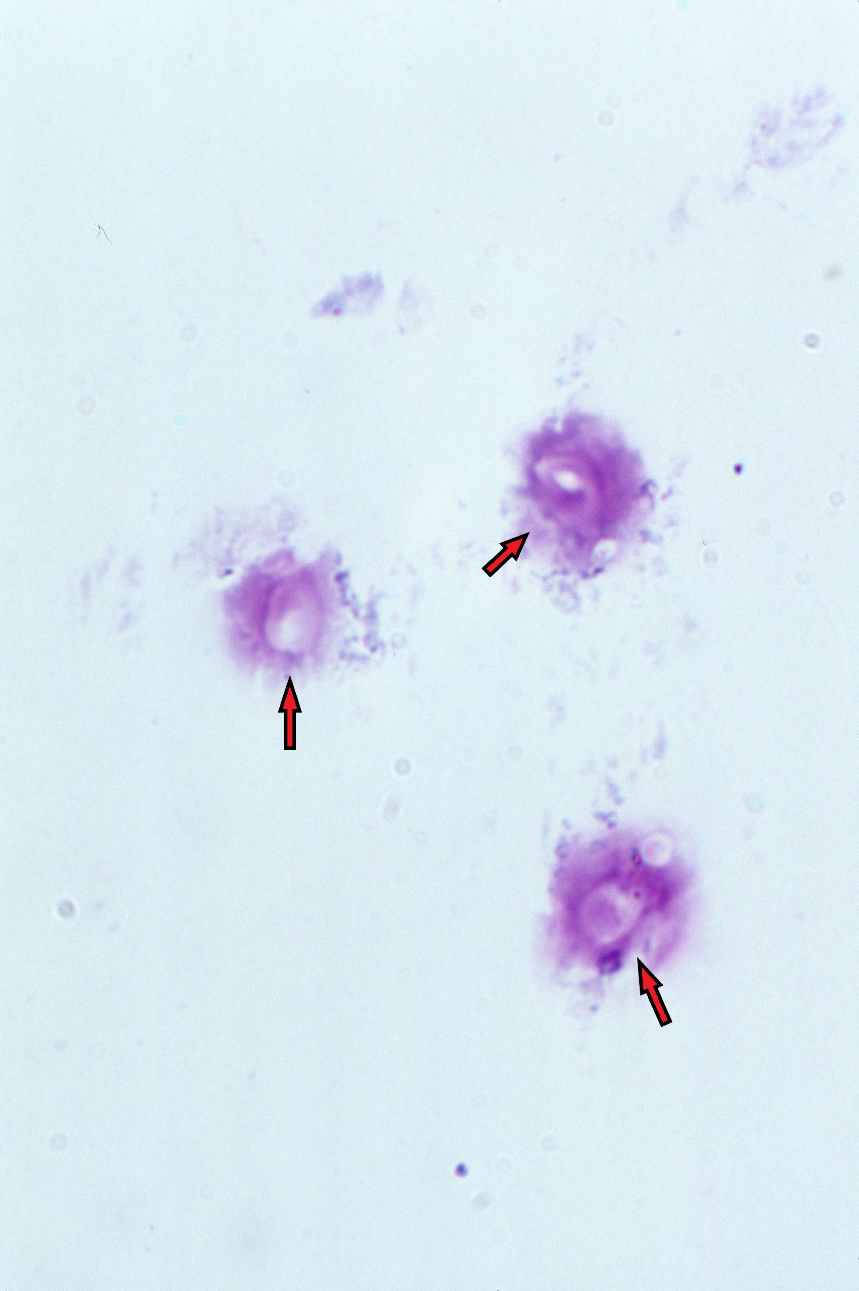
- MAG 100X Red arrows pointing to yeast cells
-
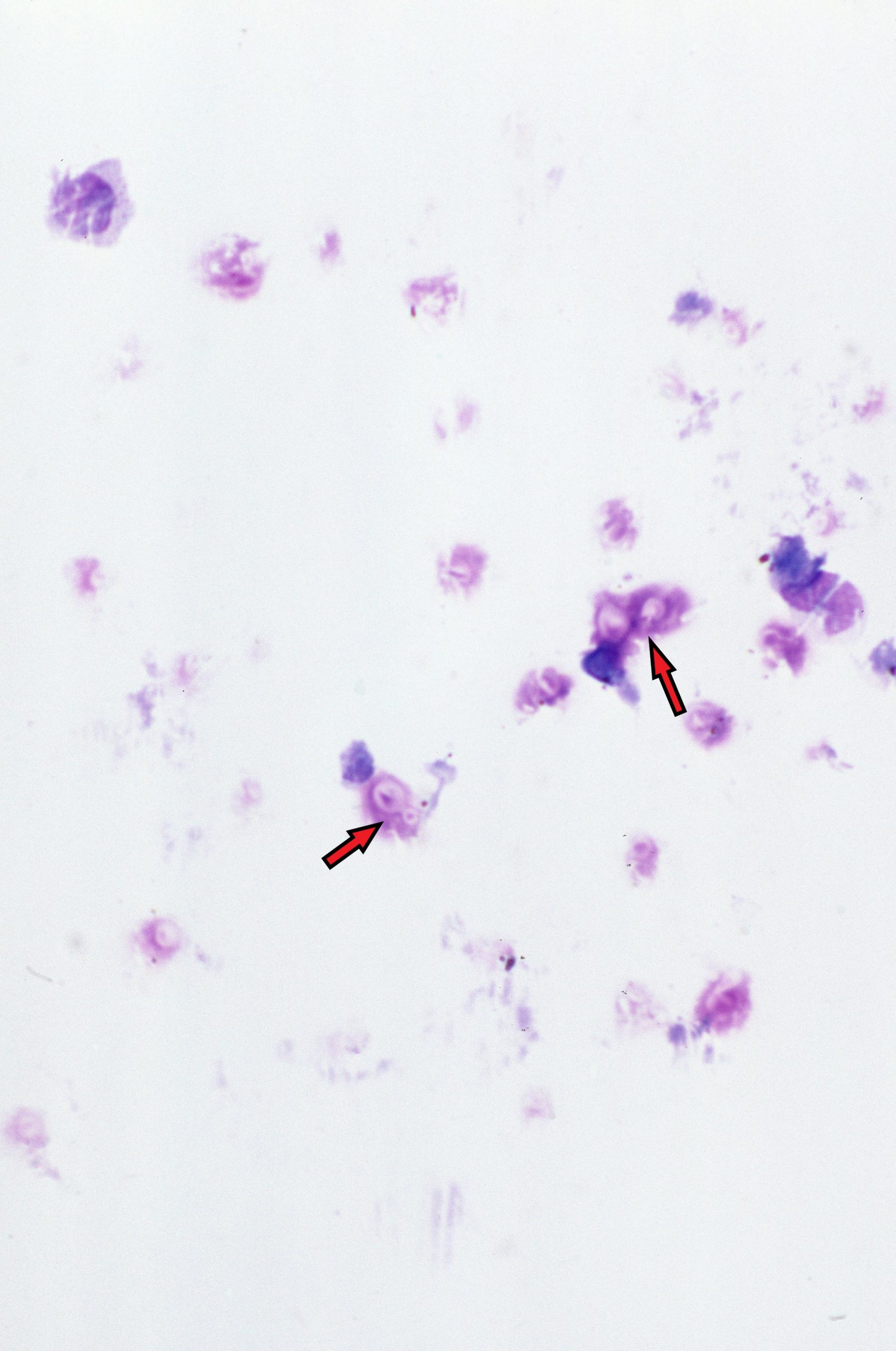
- MAG 40X Bottom red arrow points to a budding yeast cell. Note poorly stain yeast cells
-
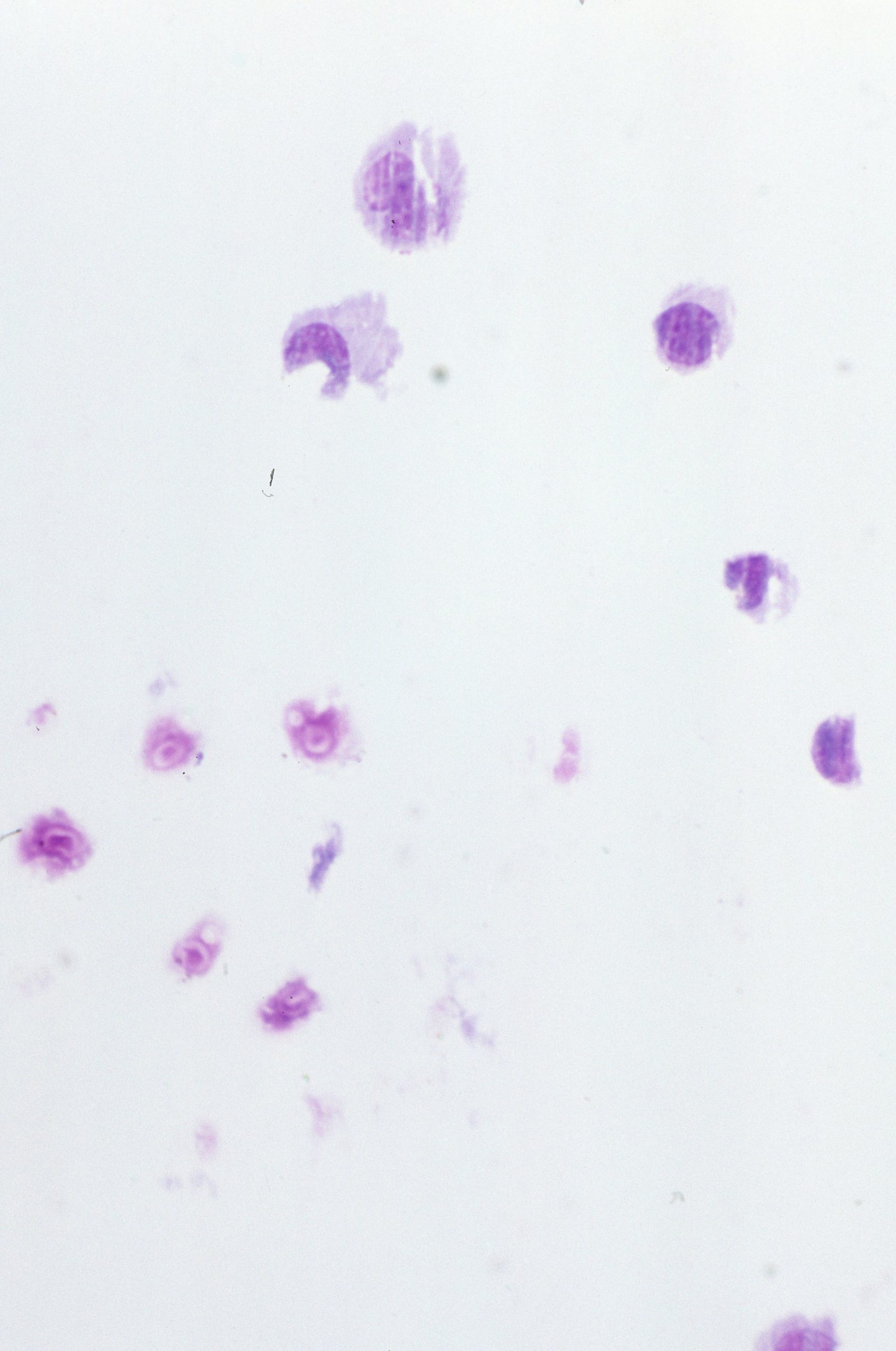
- MAX 40X
-
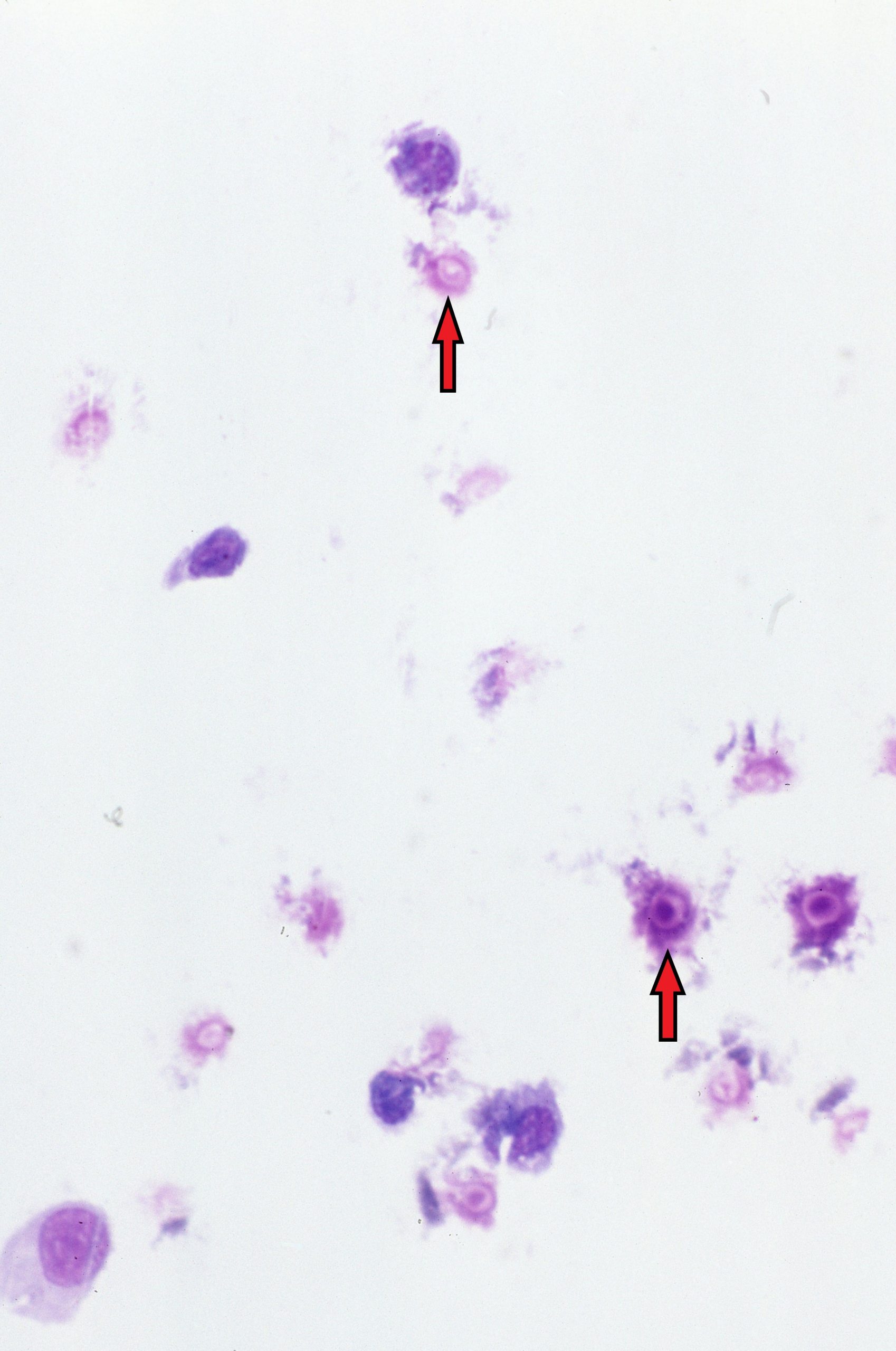
- MAG 40X
-
- MAG 100X Bottom red arrow points to a good example of a budding yeast cell. Note the Gram Positive Staining of the yeast cells